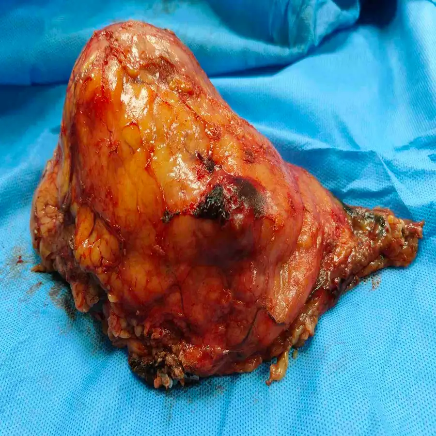
A 59-year-old male presented with abdominal pain caused by a necrotic left renal mass invading the renal sinus. Imaging also confirmed a distinct metastasis in the right adrenal gland. This aggressive bilateral presentation required immediate, highly precise surgical intervention to halt further oncological progression.
⦿ Robotic Left Radical Nephrectomy With Right Adrenalectomy
• Procedure: Advanced robotic-assisted techniques were expertly utilized for a radical nephroureterectomy and right adrenalectomy.
• Lymph Node Dissection: Seventeen retroperitoneal lymph nodes were meticulously excised and confirmed entirely free of metastasis.
• Patient Safety: Exceptional hemodynamic stability was maintained throughout, achieving a remarkably minimal blood loss of 200ml.
• Cancer Clearance: Final pathology confirmed clear margins across all tissues, ensuring complete surgical tumor eradication.
⦿ Margin-Free Clearance And Swift Recovery
The patient experienced a swift recovery, with the surgical drain and catheter successfully removed by postoperative day two. Final histopathology confirmed clear cell renal cell carcinoma with completely negative surgical margins and zero lymph node spread. He was discharged in a fully stable condition, equipped with a comprehensive follow-up endocrinology and oncology care plan.
Managing a complex renal mass alongside a contralateral adrenal metastasis demands absolute surgical precision. By utilizing an advanced robotic approach, we successfully achieved clear oncological margins while simultaneously ensuring a remarkably safe, rapid postoperative recovery.
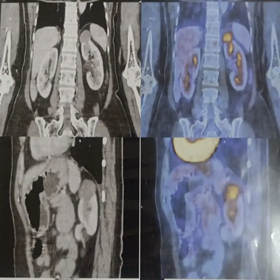
A 71-year-old female presented with left flank pain and a high-risk diagnosis of a renal mass in the superior pole of the left kidney. Diagnostic imaging identified a neoplastic lesion, necessitating urgent surgical intervention to prevent further disease progression. The patient’s condition required precise management to address the localized mass while preserving surrounding healthy renal tissue.
⦿ Robotic Left Partial Nephrectomy
• Procedure: Robotic-assisted left partial nephrectomy was successfully executed using standard multi-port access techniques.
• Complex Anatomy: The procedure involved the careful excision of a superior pole renal mass alongside an adjacent cyst.
• Safety: Precise surgical technique resulted in minimal blood loss of only 200 ml during the operation.
• Cancer Clearance: Post-operative analysis confirmed complete tumor removal with all surgical margins negative for invasive carcinoma.
⦿ Successful Recovery and Pathologic Confirmation
Postoperative recovery was uneventful, with the patient remaining hemodynamically stable and ambulatory immediately following the procedure. Final pathology confirmed a pT1a papillary renal cell carcinoma with negative regional lymph nodes and clear surgical margins. This excellent outcome allowed for a smooth transition to discharge with stable, controlled blood sugar levels and vital signs.
In this complex case, our robotic approach allowed for extreme precision, ensuring complete oncological clearance while preserving maximum renal function. By meticulously managing the resection margins, we provided the best possible long-term outlook for the patient and a swift return to daily life.
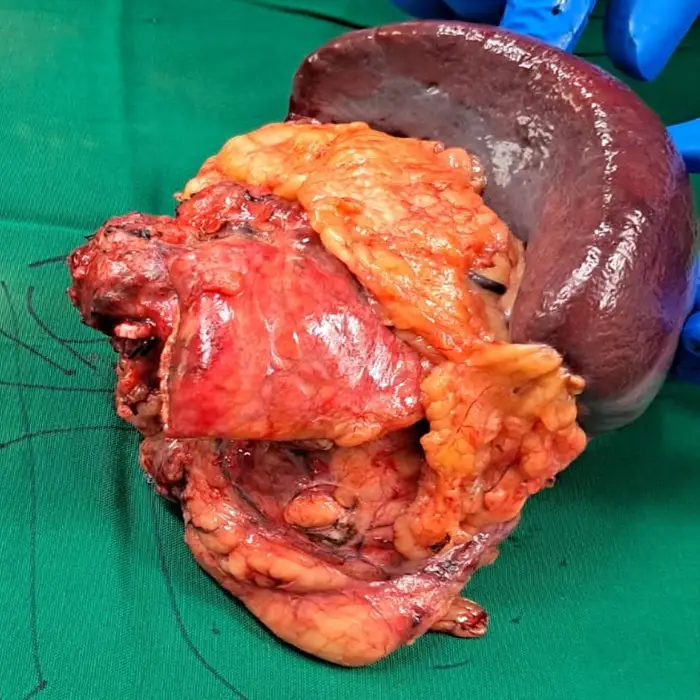
A 37-year-old female presented with left flank pain, hematuria, and marked weight loss. Imaging revealed a recurrent adrenocortical carcinoma encasing the left renal vein and infiltrating the adjacent renal parenchyma. This aggressive mass featured a complex tumor thrombus, necessitating immediate and highly precise surgical management.
⦿ Left Radical Nephrectomy with Distal Pancreatosplenectomy
• Procedure: An exploratory laparotomy achieved complete en bloc excision of the massive suprarenal tumor.
• Complex Lymphadenectomy: Eleven regional and paraaortic lymph nodes were meticulously dissected and proven negative for metastasis.
• Surgical Safety: The complex procedure maintained excellent hemodynamic stability with an exceptionally low blood loss.
• Cancer Clearance: Final pathology confirmed that all major surgical margins were completely free of malignant tissue.
⦿ Rapid Recovery And Complete Resection
Following this extensive procedure, the patient experienced an uneventful recovery and rapidly regained ambulatory function. Final pathology confirmed a complete oncological resection, demonstrating absolutely negative surgical margins across all critical tissues. Furthermore, thorough evaluations revealed zero metastatic spread across eleven dissected lymph nodes. The patient was safely discharged with stable hemodynamics and excellent functional recovery.
Managing this recurrent adrenocortical carcinoma required extreme precision to safely navigate the extensively compromised renal vasculature. By leveraging an advanced en bloc resection approach, we successfully secured completely negative surgical margins. This meticulous intervention ultimately provides our patient with the strongest possibility for long-term survival.
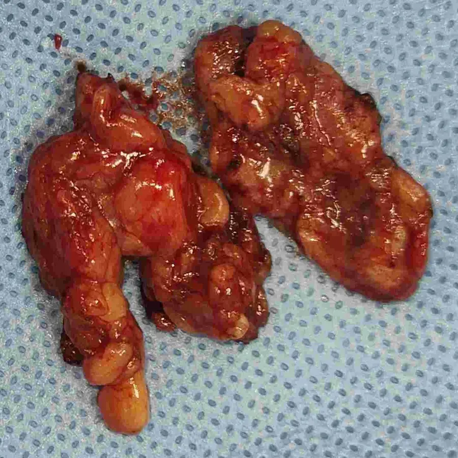
A 59-year-old female presented with persistent abdominal pain and urinary frequency, complicated by a large, heterogeneous right renal mass and ureteric calculus. Imaging suggested aggressive neoplastic potential with concurrent nodal involvement, necessitating urgent surgical intervention to address both the primary tumor and the associated symptomatic obstruction.
⦿ Surgical Nephrectomy and Excision
• Procedure: A diagnostic laparoscopy was combined with radical nephrectomy and retroperitoneal lymph node dissection.
• Anatomical Precision: The surgical team expertly navigated complex retroperitoneal anatomy to successfully excise the renal mass.
• Safety: The procedure was executed with minimal blood loss and zero requirement for blood transfusions.
• Cancer Clearance: Pathological analysis confirmed complete tumor removal with all surgical margins verified as negative.
⦿ Stable Recovery and Confirmed Diagnosis
Postoperatively, the patient remained hemodynamically stable and recovered uneventfully, demonstrating excellent tolerance to the procedure. Final histopathology identified the mass as a renal oncocytoma, with no evidence of malignancy in the analyzed lymph nodes or surgical margins. The patient was discharged in good condition, achieving a successful functional recovery.
This case highlights the critical importance of definitive surgical management when navigating challenging renal diagnostics. By employing a precise, radical approach, we effectively cleared the tumor and confirmed a favorable diagnosis, providing the patient with the best possible path to long-term health and stability.
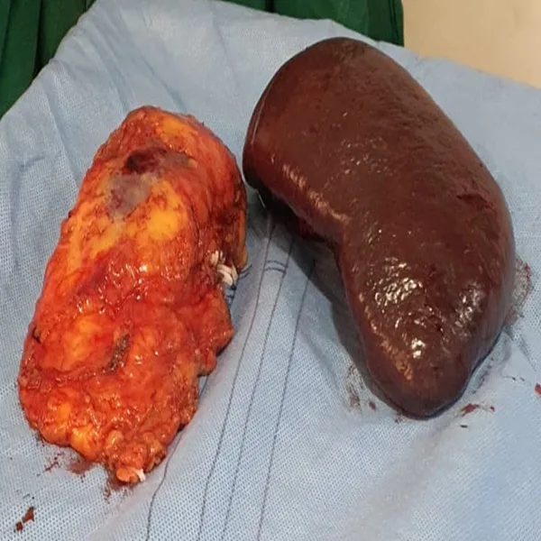
A 57-year-old male presented with a recurrent renal mass following a prior partial nephrectomy for clear cell renal cell carcinoma. The case was complicated by concurrent non-Hodgkin’s lymphoma and significant splenomegaly, necessitating a comprehensive, multidisciplinary surgical approach to manage these overlapping clinical and anatomical challenges effectively.
⦿ Robotic-Assisted Nephrectomy and Splenectomy
• Procedure: A robotic-assisted right radical nephrectomy and splenectomy was successfully executed using standard port placement.
• Complex Anatomy: The surgery successfully overcame dense adhesions between the gastric fundus and the splenic hilum.
• Safety: The procedure was completed with controlled blood loss, maintaining the patient's hemodynamic stability throughout surgery.
• Cancer Clearance: Pathological examination confirmed complete resection of the renal carcinoma with all surgical margins being negative.
⦿ Successful Recovery and Pathologic Confirmation
The patient remained hemodynamically stable postoperatively and was mobilized effectively following the robotic intervention. Final pathology confirmed a pT1a renal cell carcinoma with clear, margin-free resection of the renal mass and successful removal of the spleen. The patient achieved a stable recovery, allowing for a successful and timely hospital discharge.
Managing recurrent renal malignancy requires both clinical precision and the versatility of robotic platforms to handle complex adhesions. By utilizing this advanced approach, we successfully achieved complete oncological clearance while ensuring the patient’s safety and facilitating a smooth, functional recovery for long-term health.
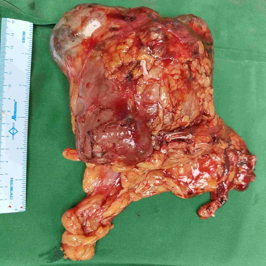
A 64-year-old male presented with persistent pain and weight loss, leading to the diagnosis of a high-complexity left renal solid cystic mass. The lesion was identified as a potentially aggressive renal cell carcinoma, showing heterogeneous enhancement and requiring urgent surgical intervention to prevent further disease progression.
⦿ Laparoscopic Radical Nephrectomy and Retroperitoneal Lymph Node Dissection
• Procedure: The surgery utilized a comprehensive laparoscopic radical nephrectomy and retroperitoneal lymph node dissection.
• Complex Anatomy: The surgical team navigated high-complexity tumor geography near the collecting system with remarkable anatomical accuracy.
• Patient Safety: The procedure was executed with exceptional precision, resulting in minimal blood loss of 150 ml.
• Cancer Clearance: Histopathological analysis confirmed the complete removal of the tumor with all surgical margins negative.
⦿ Successful Recovery and Oncological Clearance
Postoperatively, the patient exhibited an uneventful recovery with stable hemodynamics and regained mobility shortly after the procedure. Pathology confirmed Clear Cell Renal Cell Carcinoma with tumor extent limited to the kidney and negative surgical margins. The patient was discharged in a stable condition, marking a highly successful oncological outcome for this complex case.
By utilizing advanced laparoscopic techniques, we were able to safely remove this high-complexity renal mass while ensuring oncological integrity. This precise approach provided the patient with the best possible chance for a complete cure and a favorable long-term survival outlook.
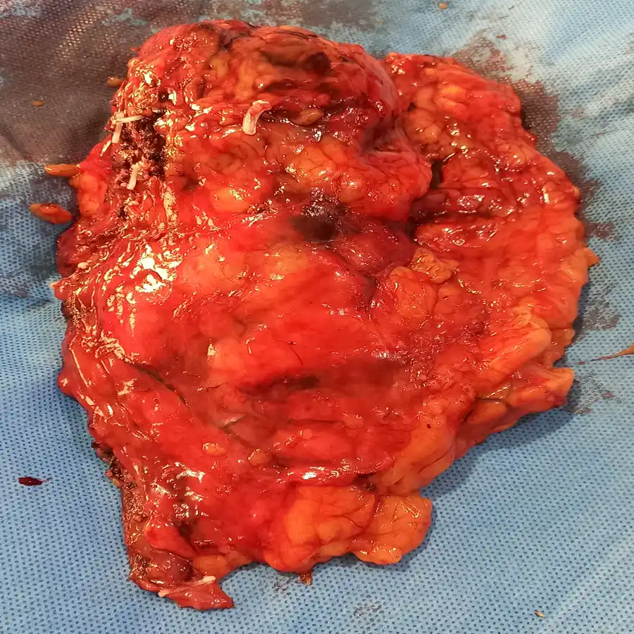
A 62-year-old male presented with abdominal pain and a significant 8cm left renal mass. Imaging confirmed a complex tumor extending into the renal pelvis and adhering to the diaphragm, requiring specialized surgical intervention.
⦿ Robotic Left Extended Radical Nephrectomy
• Procedure: A trans-peritoneal approach was utilized with 5 robotic ports and a Pfannenstiel incision.
• Anatomical Complexity: The tumor involved the mid and upper renal poles with enlarged para-aortic lymph nodes.
• Safety: The procedure was executed with exceptional safety, resulting in minimal blood loss of 100ml.
• Completeness: The entire tumor specimen was successfully retrieved and sent for histopathological analysis to ensure clearance.
⦿ Stable Recovery and Discharge
The patient experienced an uneventful postoperative recovery, maintaining stable hemodynamics and controlled pain levels throughout his hospital stay. He remained ambulatory following the procedure and was discharged with a clean surgical wound and well-managed blood sugar levels. This successful robotic approach provided the patient with the most favorable path toward long-term recovery and health.
By leveraging the precision of robotic-assisted technology, we successfully navigated the complex adhesions involving the diaphragm to ensure a complete oncological resection. This approach allowed for a minimally invasive recovery, offering the patient the best possible chance at long-term cure.
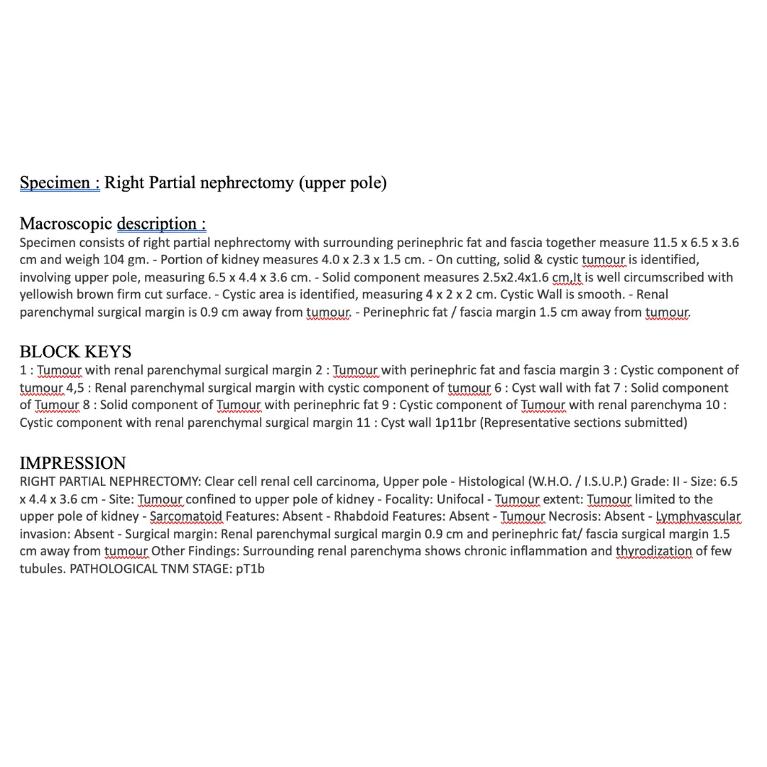
This sixty-three-year-old male patient presented with a large, exophytic right renal mass measuring fifty-eight millimeters. The complexity was heightened because he had previously received definitive chemo-radiation therapy for esophageal carcinoma. Managing this new cT2N0 upper pole tumor required precise surgical intervention.
⦿ Robotic Right Partial Nephrectomy
• Procedure: Dr. Swati Shah successfully executed a partial nephrectomy keeping a half-centimeter circumferential margin.
• Complex Anatomy: Careful adhesiolysis and pyeloplasty were required to manage the challenging residual kidney anatomy.
• Surgical Safety: Hemostasis was meticulously ensured after a short warm ischemia time of thirty minutes.
• Cancer Clearance: The large solid-cystic upper pole tumor was completely removed without gross adjacent invasion.
⦿ Successful Recovery And Preservation
The patient experienced an extremely smooth post-operative recovery, achieving stable hemodynamics and an ambulatory status very quickly. Intraoperative findings confirmed the precise removal of the mass while maintaining an adequate half-centimeter surgical margin. Ultimately, the patient achieved excellent functional stability and was discharged safely on medications.
Navigating a new renal tumor in a patient with a prior esophageal malignancy requires absolute surgical precision. By utilizing an advanced partial nephrectomy technique, we successfully eliminated the cancer while preserving vital kidney function.
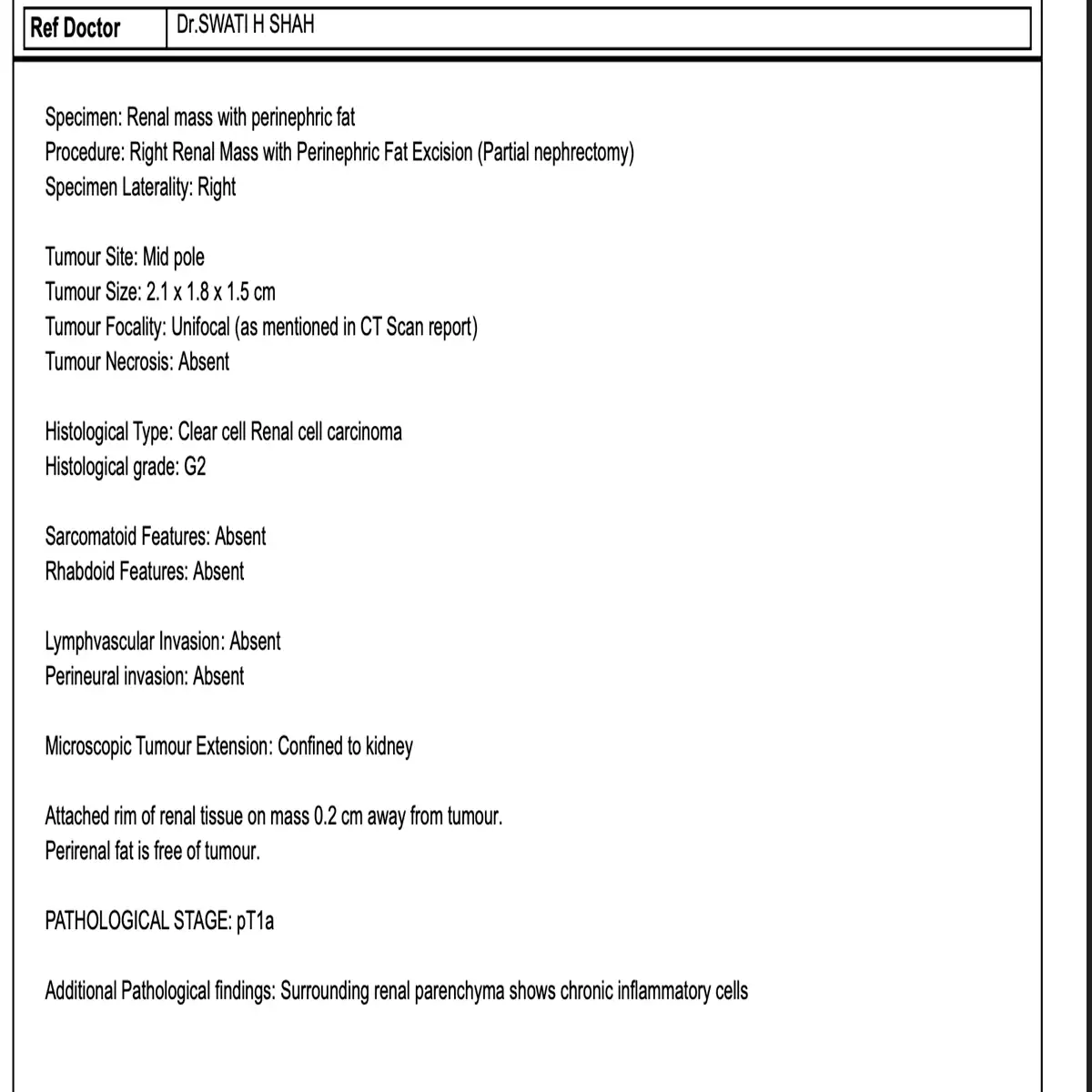
A 37-year-old male was incidentally found to have a right renal mass during an evaluation for peptic disease. Imaging revealed a suspicious 24 millimeter lesion located directly in the midpole of the right kidney. Surgical intervention was critically required to safely remove this tumor while maximizing the preservation of healthy kidney function.
⦿ Robotic Right Partial Nephrectomy
• Surgical Approach: A five-port trans-peritoneal method safely exposed the right midpole renal mass.
• Vascular Control: The renal vessels were precisely clamped to achieve a safe 37-minute warm ischemia time.
• Patient Safety: Complete hemostasis was carefully ensured, leading to a remarkably smooth procedure without postoperative complications.
• Tumor Clearance: Final pathology strictly confirmed a totally excised pT1a clear cell renal cell carcinoma with complete margin-free status.
⦿ Exceptional Recovery and Complete Cancer Clearance
The patient experienced an excellent postoperative recovery and was safely discharged in a hemodynamically stable, highly ambulatory condition. Final pathology results completely confirmed localized stage pT1a disease with absolutely free surgical margins and zero lymphovascular invasion. Ultimately, the patient retained exceptional residual kidney function and successfully returned to normal physical activities.
By utilizing an advanced robotic approach, I successfully excised this incidental renal mass while perfectly preserving the vital surrounding kidney tissue. This high degree of technical precision safely ensures excellent oncological clearance, providing the patient with the absolute best opportunity for a long-term, cancer-free future.
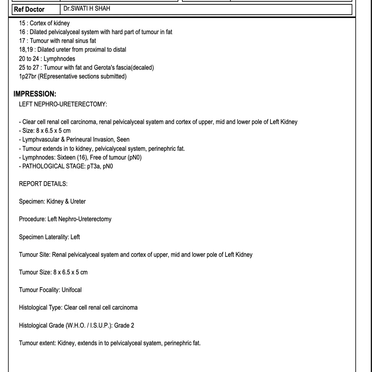
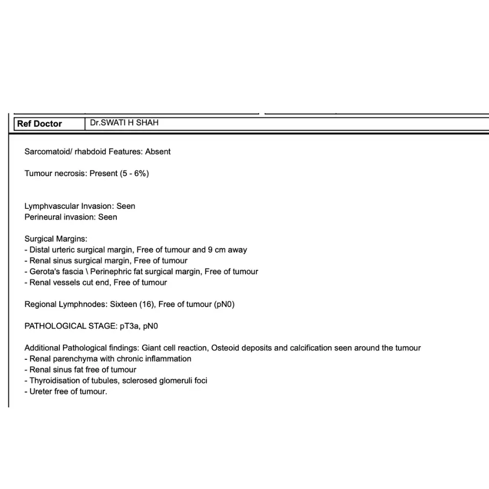
A 59-year-old female presented with a significant history of left flank pain and hematuria, complicating a diagnosis of a large renal pelvic mass. Advanced imaging confirmed a complex tumor involving the kidney and pelvicalyceal system, requiring urgent intervention to manage this aggressive malignancy and prevent further systemic progression.
⦿ Minimally Invasive Robotic Nephroureterectomy
• Procedure: A minimally invasive robotic radical nephroureterectomy was executed using a five-port transperitoneal surgical approach.
• Lymph Node Management: Sixteen regional lymph nodes were meticulously harvested and confirmed to be entirely free of malignancy.
• Safety and Precision: The robotic intervention facilitated precise vascular control and minimal surgical trauma throughout the complex removal process.
• Cancer Clearance: Pathological analysis confirmed complete tumor removal with clear surgical margins, ensuring an optimal oncological outcome.
⦿ Successful Recovery and Pathological Clearance
Postoperatively, the patient remained hemodynamically stable and exhibited a swift, uncomplicated recovery. Final histopathology revealed clear cell renal cell carcinoma with no nodal involvement, confirming the success of the surgical approach. She was discharged in excellent condition, demonstrating the efficacy of robotic-assisted surgery in achieving comprehensive cancer clearance and rapid functional stabilization.
In this complex case, leveraging robotic technology was instrumental in achieving a radical yet precise resection. By meticulously clearing all regional nodes and ensuring negative margins, we provided the best possible foundation for a long-term cure and superior patient recovery.
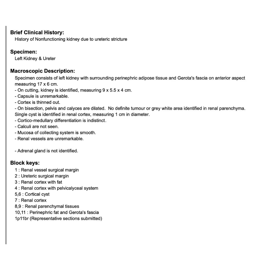
A 52-year-old male presented with a year-long history of intermittent left flank pain and reduced urine output. Clinical investigations confirmed a nonfunctioning kidney secondary to an upper ureteric stricture, complicated by chronic pyelonephritis and high-grade fevers. The patient’s condition necessitated surgical intervention to address the severe renal dysfunction.
⦿ Robotic Left Nephrectomy
• Procedure: A robotic-assisted left nephrectomy was successfully completed via a 5-port trans-peritoneal approach.
• Anatomical Challenge: The procedure required meticulous medial reflection of the colon to safely expose the renal hilum.
• Safety Profile: The surgical approach prioritized minimal blood loss and maximized patient safety throughout the intervention.
• Tissue Removal: The procedure achieved the complete and precise removal of the damaged renal specimen.
⦿ Successful Recovery and Pathological Resolution
The patient remained hemodynamically stable post-operatively and was ambulatory shortly after the surgery. Histopathology confirmed the diagnosis of chronic pyelonephritis, characterized by interstitial inflammatory cells and dilated calyces. The patient achieved an uneventful recovery and was discharged in satisfactory condition with stable vital parameters and effective pain control.
In this case, the Da Vinci Xi system provided the superior visualization necessary to navigate the complex inflammatory changes safely. By opting for a minimally invasive robotic approach, we ensured a precise removal of the damaged tissue, facilitating a faster recovery for the patient.
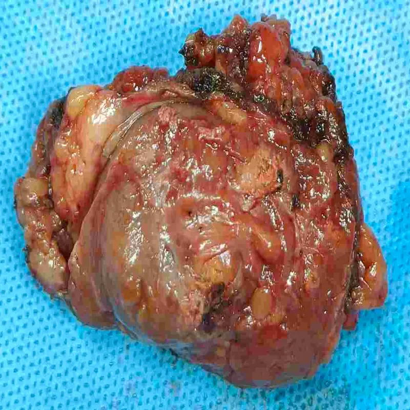
A 66-year-old male presented with a symptomatic 3.5 cm renal mass and bilateral inguinal hernias. Imaging suggested potential renal cell carcinoma, necessitating precise surgical intervention to preserve renal function while ensuring complete tumor eradication. The combined presentation required a sophisticated, multi-disciplinary approach to address both the oncological and abdominal concerns.
⦿ Robotic Right Partial Nephrectomy and Open Bilateral Inguinal Hernioplasty
• Procedure: A robotic trans-peritoneal approach was utilized for the partial nephrectomy and hernia repair.
• Anatomical Complexity: The surgery successfully addressed a lower-pole renal mass alongside bilateral indirect inguinal hernias.
• Patient Safety: The procedure was executed with exceptional safety, resulting in minimal blood loss of 200 ml.
• Cancer Clearance: Postoperative histopathology confirmed an oncocytoma with all surgical margins negative for invasive tumor cells.
⦿ Seamless Recovery and Excellent Pathological Success
The patient demonstrated an uneventful postoperative recovery and remained hemodynamically stable throughout his hospital stay. Final pathology confirmed a complete excision of the oncocytoma with clear margins, negating the need for further oncological intervention. He was discharged in a stable, ambulatory condition, having successfully resolved both the renal neoplasm and bilateral hernias.
By utilizing the Da Vinci robotic system, we were able to perform a highly precise partial nephrectomy that prioritized renal preservation. This approach provided the best possible oncological outcome for the patient, ensuring a swift recovery and long-term functional stability.
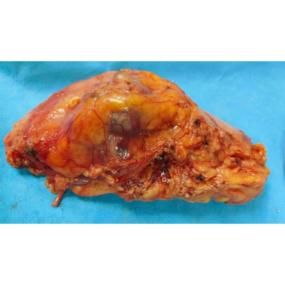
A 51-year-old male presented with significant abdominal discomfort, leading to the discovery of a large right renal lesion. Imaging revealed an intermediate-to-high complexity mass measuring over eight centimeters, heavily abutting critical structures like the duodenum.
⦿ Robotic Radical Nephrectomy
Dr. Swati Shah performed a highly precise Robotic Radical Nephrectomy to safely excise the extensive right-sided tumor. Utilizing advanced robotic articulation, the surgical team meticulously dissected the mass away from the adherent duodenum and safely secured the right renal vasculature.
• Procedure: Robotic-assisted right radical nephroureterectomy was performed using a standard six-port approach.
• Complex Anatomy: The procedure required meticulous adhesiolysis due to the mass abutting the duodenum.
• Safety: The surgery was highly efficient, resulting in minimal blood loss of only 200 ml.
• Cancer Clearance: Pathological analysis confirmed complete tumor removal with all surgical margins negative for carcinoma.
⦿ Complete Oncological Clearance And Swift Recovery
The patient experienced an uneventful postoperative recovery and was safely discharged with stable hemodynamics. Final histopathology revealed a grade two clear cell renal cell carcinoma, but importantly, all surgical margins were definitively negative for invasive disease. He successfully resumed normal ambulatory activities, achieving an excellent functional outcome and optimal oncological prognosis.
"By leveraging robotic precision, we safely navigated the complex adhesions surrounding the duodenum and secured the renal hilum. This minimally invasive approach ensured completely negative margins for a large tumor, providing the patient with an optimal pathway for a definitive cure."
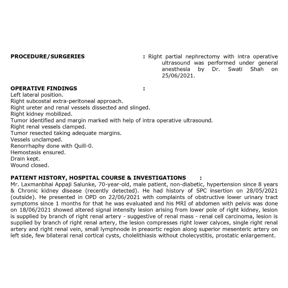
A 70-year-old male presented with a significant right renal mass and a complete urethral stricture, complicating his management. His medical history included chronic kidney disease and hypertension, necessitating a highly specialized approach to address both the oncological challenge and the obstructive urinary symptoms.
⦿ Right Partial Nephrectomy
• Procedure: A right partial nephrectomy was executed using an extra-peritoneal approach with specialized intra-operative ultrasound guidance.
• Anatomical Complexity: The surgeon navigated complex anatomy, specifically managing the renal mass compressing the right lower calyces.
• Safety: Hemostasis was meticulously ensured after resection, resulting in a stable and uneventful surgical procedure.
• Cancer Clearance: The tumor was successfully resected with adequate margins to ensure comprehensive oncological control.
⦿ Successful Recovery and Stable Discharge
The patient’s post-operative recovery was smooth, uneventful, and characterized by hemodynamic stability throughout his hospital stay. Following the successful partial nephrectomy, he was discharged in a stable condition with appropriate catheter care plans in place. This approach allowed for effective tumor management despite the patient's underlying chronic kidney disease.
Managing a renal mass alongside a complete urethral stricture in a patient with chronic kidney disease requires absolute precision to preserve renal function. By utilizing an extra-peritoneal approach, we achieved complete tumor resection, offering the patient the best possible oncological outcome.
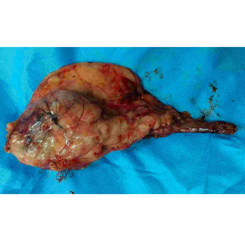
A female patient presented with a completely non-functional, atrophic right kidney complicated by a thickened ureter. The surgical challenge was significantly increased by dense, severe adhesions binding the kidney to the gonadal and external iliac vessels. Enlarged paracaval lymph nodes also required careful clinical attention.
⦿ Robotic Right Nephrectomy with Adhesiolysis
• Procedure: A complex robotic right nephroureterectomy and adhesiolysis were executed with exceptional surgical precision.
• Complex Anatomy: The team successfully navigated dense anatomical adhesions surrounding the external iliac and gonadal vessels.
• Blood Loss: Patient safety was prioritized, resulting in an exceptionally minimal estimated blood loss of 50ml.
• Cancer Clearance: Complete removal of the atrophic kidney, ureter, and enlarged paracaval lymph nodes was achieved.
⦿ Successful Recovery and Tissue Removal
Following the surgery, the patient achieved excellent hemostasis and remained completely hemodynamically stable throughout the closure. The complete right nephroureterectomy specimen and paracaval lymph nodes were successfully retrieved and sent for histopathological evaluation. Thanks to the minimally invasive robotic approach, the patient experienced a rapid postoperative recovery with excellent long-term functional prospects.
Navigating the dense adhesions around the iliac vessels required extreme surgical precision to safely remove the non-functional kidney. The advanced robotic platform allowed us to perform this complex extraction flawlessly while preserving surrounding vital structures, ensuring an outstanding recovery for our patient.
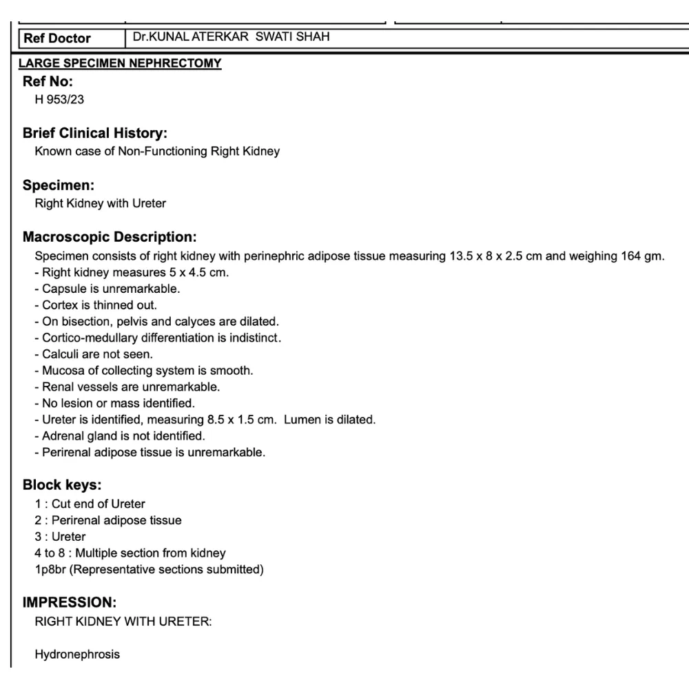
A 57-year-old female presented with persistent right flank pain, lower urinary tract symptoms, and vomiting lasting 15 days. Diagnostic imaging and evaluation confirmed a non-functioning right kidney. This condition necessitated surgical intervention to resolve the patient’s discomfort and address the underlying chronic inflammatory pathology effectively.
⦿ Robotic Right Nephrectomy
• Procedure: The surgery utilized a robotic-assisted approach for precise removal of the affected kidney.
• Complex Anatomy: The procedure successfully managed a small, atrophied kidney with associated hydronephrosis and hydroureter.
• Safety: Hemostasis was meticulously achieved throughout the surgery to ensure high levels of patient safety.
• Cancer Clearance: Histopathological examination confirmed the complete removal of the diseased specimen with no malignancy identified.
⦿ Stable Recovery and Pathological Resolution
Following the robotic procedure, the patient remained hemodynamically stable and demonstrated a smooth post-operative recovery. Final pathology reports revealed chronic pyelonephritis, confirming the absence of malignancy and successful clearance of the pathology. The patient was discharged in good condition, showing positive functional improvement and achieving full recovery.
By utilizing robotic technology, we achieved superior precision in managing this complex, non-functioning kidney. This minimally invasive approach allowed for a swift, stable recovery and provided the patient with the best possible long-term outcome, effectively resolving her chronic symptoms and improving her quality of life.
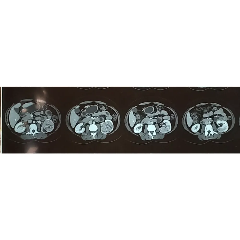
A 46-year-old female presented with hematuria and was subsequently diagnosed with a substantial left renal mass. Imaging revealed a large 9×6 cm lesion extending into the perinephric fat and pelvicalyceal system. The tumor’s size and complex location demanded immediate and highly precise surgical intervention.
⦿ Robotic Left Radical Nephrectomy
• Procedure: An advanced robotic radical nephrectomy seamlessly excised the compromised kidney and affected tissue.
• Complex Anatomy: The intricate retroperitoneal lymph node dissection was executed with exceptional robotic magnification.
• Patient Safety: Strict hemostasis ensured optimal patient safety with a minimal blood loss of 50 ml.
• Cancer Clearance: Complete en bloc removal of the tumor was achieved without any extra-gerotal spread.
⦿ Rapid Recovery and Disease Control
The patient experienced an uneventful postoperative recovery and was ambulatory with stable hemodynamics shortly after surgery. Final histopathology confirmed clear cell renal cell carcinoma, completely resected alongside the para-aortic lymph nodes. She was safely discharged on postoperative day three, ready to resume normal diet and activities.
By utilizing advanced robotic techniques for this large tumor, we achieved precise vascular control and complete oncological clearance. This minimally invasive approach drastically reduced recovery time while providing the patient with the best possible chance for long-term survival and excellent quality of life.
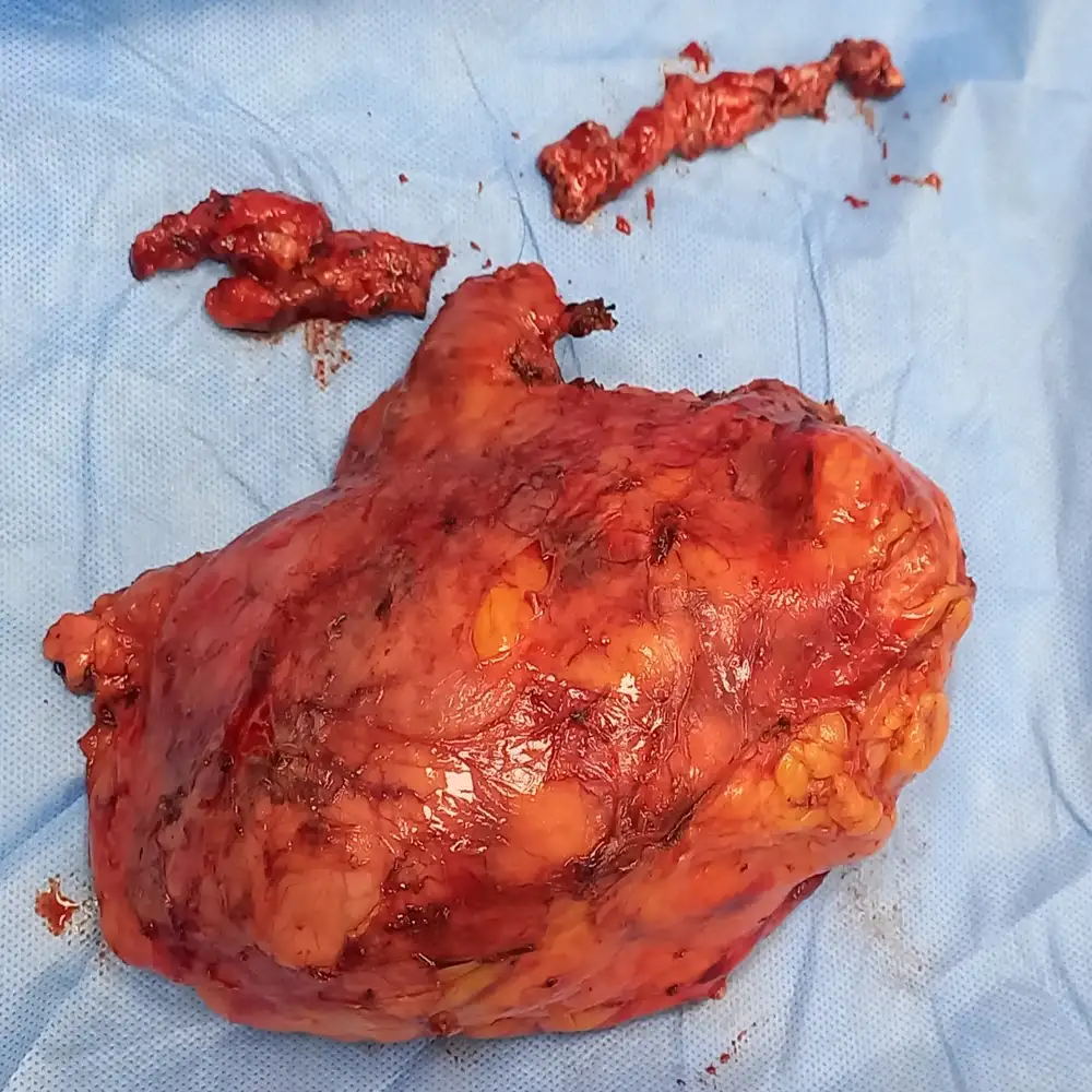
A 59-year-old male presented with significant hematuria and urinary retention caused by a large, aggressive left renal cell carcinoma. Imaging revealed high-risk tumor thrombus extending into the infrahepatic inferior vena cava, alongside retroperitoneal lymphadenopathy and bladder clot obstruction.
⦿ Exploratory Laparotomy and Radical Nephrectomy
• Procedure: A radical nephrectomy and retroperitoneal tumor excision were performed via a chevron subcostal incision.
• Complex Anatomy: The surgery addressed a massive renal lesion and high-level infrahepatic IVC tumor thrombus.
• Safety: Hemostasis was expertly achieved throughout the extensive resection to ensure optimal intraoperative patient safety.
• Cancer Clearance: Pathological examination confirmed complete tumor excision with negative surgical margins and no lymph node metastasis.
⦿ Exceptional Postoperative Recovery
The patient experienced an uneventful postoperative recovery and remained hemodynamically stable throughout his hospital stay. Final pathology indicated pT3b, pN0 staging, confirming successful radical resection without lymph node involvement. He achieved controlled blood sugar levels and was discharged in an ambulatory, stable condition to continue his recovery at home.
Managing advanced renal cell carcinoma with level II IVC thrombus requires extreme surgical precision and careful vascular handling. By performing a complete radical resection, we successfully removed the malignancy while minimizing risks to restore the patient's long-term health prospects.
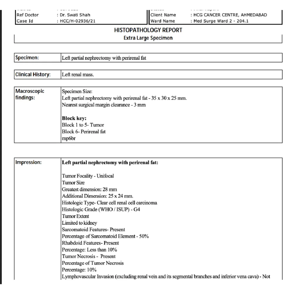
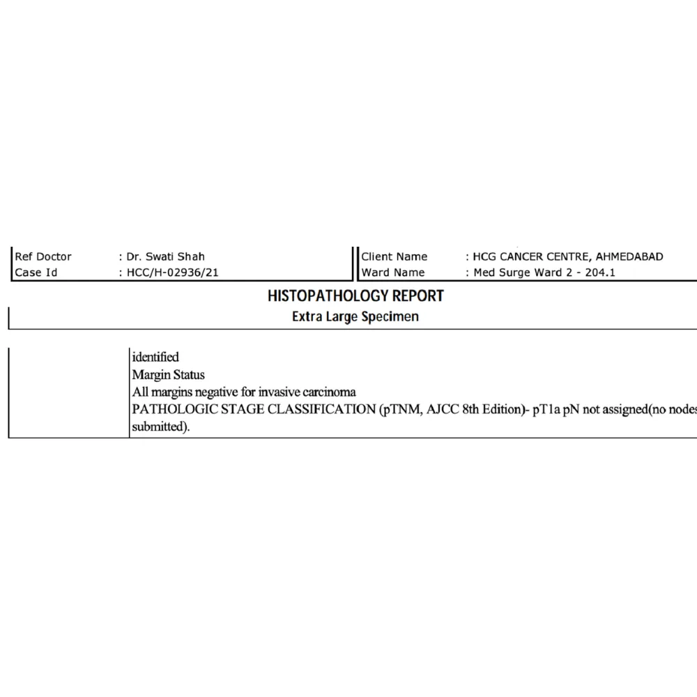
A 57-year-old male presented with fever and abdominal pain, leading to the diagnosis of a localized renal mass in the lower pole of the left kidney. Imaging confirmed a complex neoplastic lesion necessitating immediate surgical intervention to address the malignancy while preserving healthy renal function.
⦿ Robotic Left Partial Nephrectomy
• Procedure: A minimally invasive robotic-assisted partial nephrectomy was successfully executed using the Da Vinci platform.
• Anatomical Precision: The surgery involved careful medial reflection of the colon to safely expose the renal hilum.
• Safety Profile: Hemostasis was strictly ensured following a brief warm ischemia time of twenty-three minutes.
• Cancer Clearance: The procedure achieved complete tumor excision with all surgical margins confirmed as negative for carcinoma.
⦿ Successful Recovery and Pathologic Confirmation
The patient’s post-operative recovery was smooth and uneventful, with the patient remaining hemodynamically stable throughout the hospital stay. Final histopathology confirmed a clear cell renal cell carcinoma, successfully removed with negative surgical margins. The patient was discharged in stable condition, ready to resume normal activities under continued clinical surveillance.
By utilizing the robotic platform, we were able to precisely excise the tumor while effectively preserving the patient’s renal function. This targeted approach ensures the best oncological outcome, allowing us to manage aggressive renal malignancy with minimal trauma and rapid patient recovery.
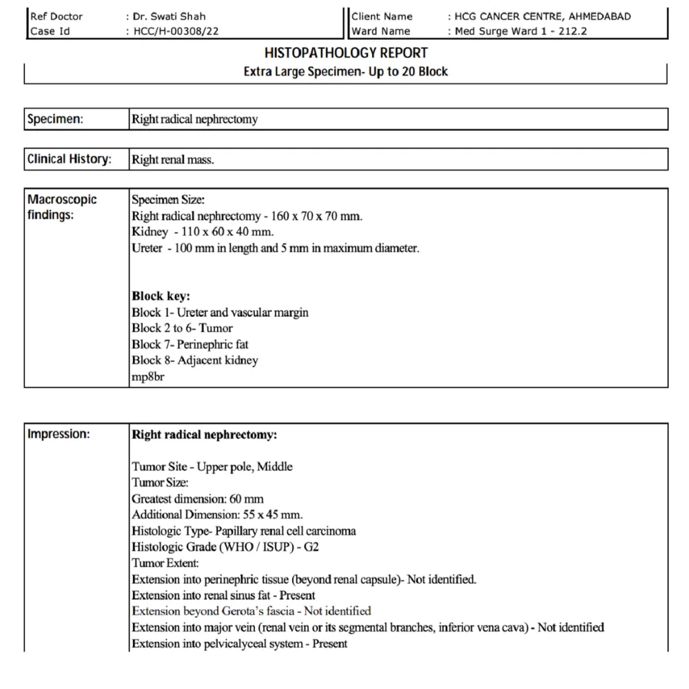
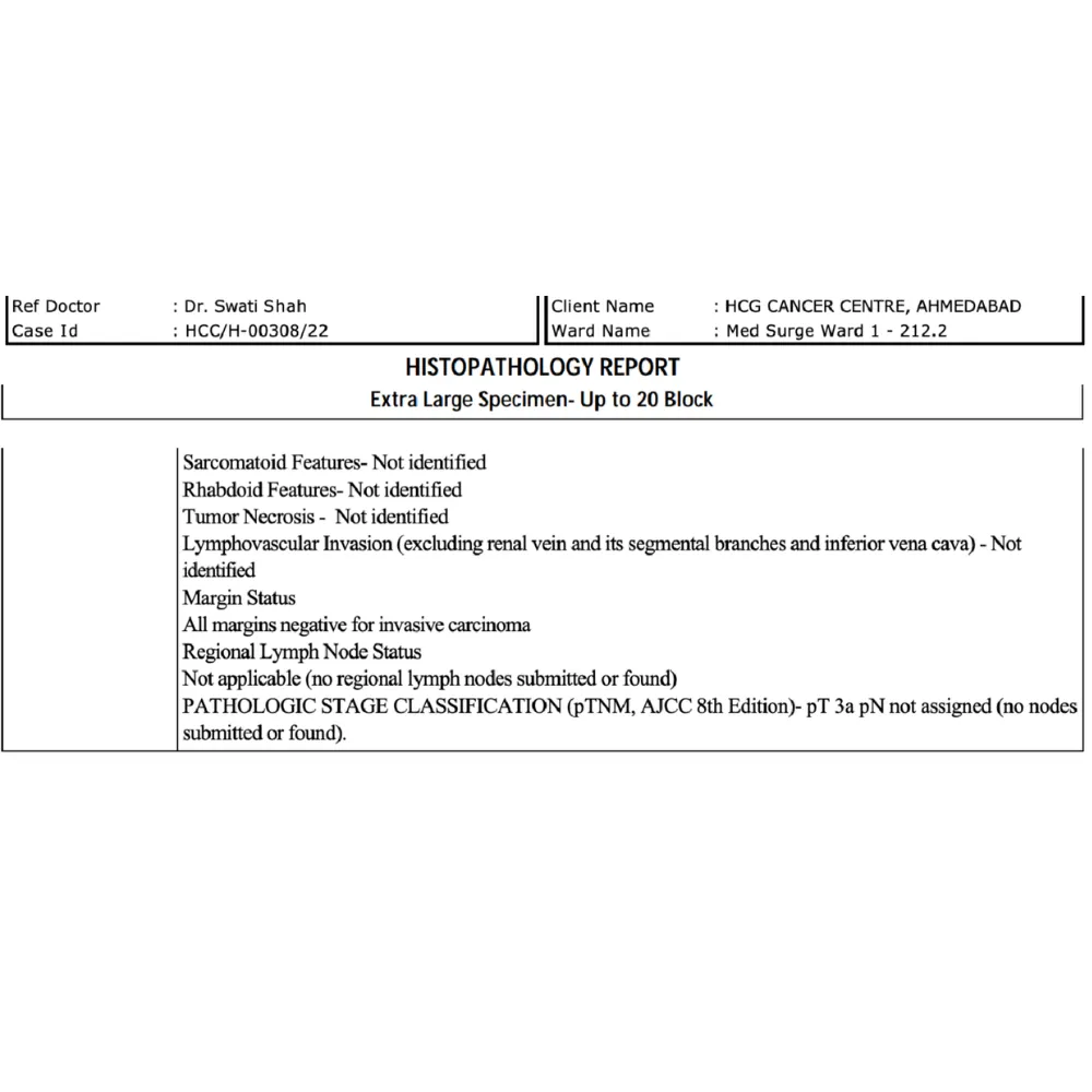
A 55-year-old female presented with a complex right renal mass requiring immediate surgical intervention. Evaluation revealed a 60 mm tumor extending into the renal sinus fat and pelvicalyceal system. This advanced presentation necessitated a highly precise oncological approach to ensure total tumor clearance.
⦿ Laparoscopic Right Radical Nephrectomy
• Procedure: A minimally invasive laparoscopic radical nephrectomy was successfully executed for optimal tumor removal.
• Anatomical Challenge: The large 60 mm tumor complexly involved both renal sinus and pelvicalyceal structures.
• Patient Safety: Advanced laparoscopic techniques significantly reduced procedural blood loss and ensured rapid postoperative recovery.
• Cancer Clearance: Pathological analysis confirmed all surgical margins were completely negative for any invasive carcinoma.
⦿ Cancer-Free With Excellent Recovery
The patient experienced an excellent postoperative recovery and remained hemodynamically stable throughout the hospital stay. Final histopathology revealed a Grade 2 papillary renal cell carcinoma with completely negative surgical margins. Through this advanced approach, the patient achieved complete cancer clearance and a rapid return to daily activities.
By utilizing a targeted laparoscopic approach for this complex 60 mm tumor, I was able to safely achieve absolute negative margins. This minimally invasive method ensured complete oncological control and provided the best foundation for long-term survival.
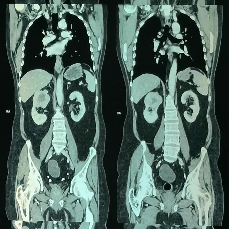
A 60-year-old male presented with urinary symptoms and was subsequently diagnosed with a bulky renal cell carcinoma. Imaging revealed a large, centrally necrotic lesion in the right kidney abutting the liver, further complicated by multiple bony metastases. This aggressive presentation required prompt and meticulous surgical intervention.
⦿ Right Radical Nephrectomy
• Procedure: An advanced radical nephrectomy was successfully executed to remove the entire affected right kidney.
• Complex Anatomy: The bulky tumor's extension toward the renal hilum required meticulous dissection and vessel ligation.
• Patient Safety: Excellent hemostasis was maintained throughout the operation, ensuring highly stable intraoperative patient vitals.
• Cancer Clearance: The diseased kidney, surrounding perinephric fat, and right adrenal gland were fully excised.
⦿ Uneventful Recovery And Discharge
Following the procedure, the patient experienced a remarkably smooth and uneventful postoperative recovery within the specialized oncology unit. Final histopathology confirmed the complete removal of an eosinophilic renal tumor through our meticulously managed surgical approach. He regained independent ambulatory function rapidly and was discharged home in a completely stable hemodynamic condition.
Managing such a large, locally advanced renal mass requires absolute anatomical precision to prevent complications. By performing a meticulous radical nephrectomy, we successfully eliminated the primary source of the disease, granting the patient the strongest possible foundation for his ongoing oncological therapies.
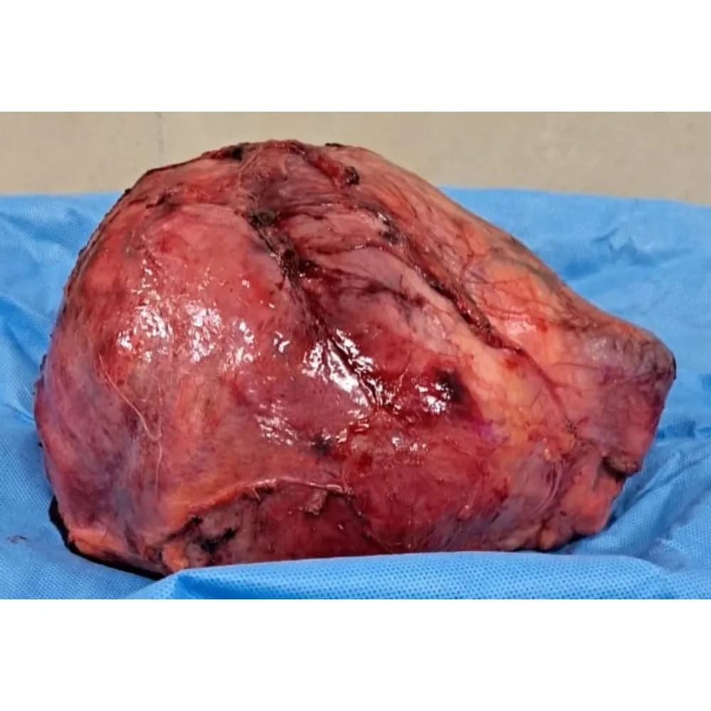
A 42-year-old male presented with a massive, aggressive right renal mass compressing the inferior vena cava, alongside profound local lymph node involvement. The aggressively growing tumor, previously causing severe cough and vomiting, demanded urgent surgical intervention to prevent further vascular invasion and systemic metastasis.
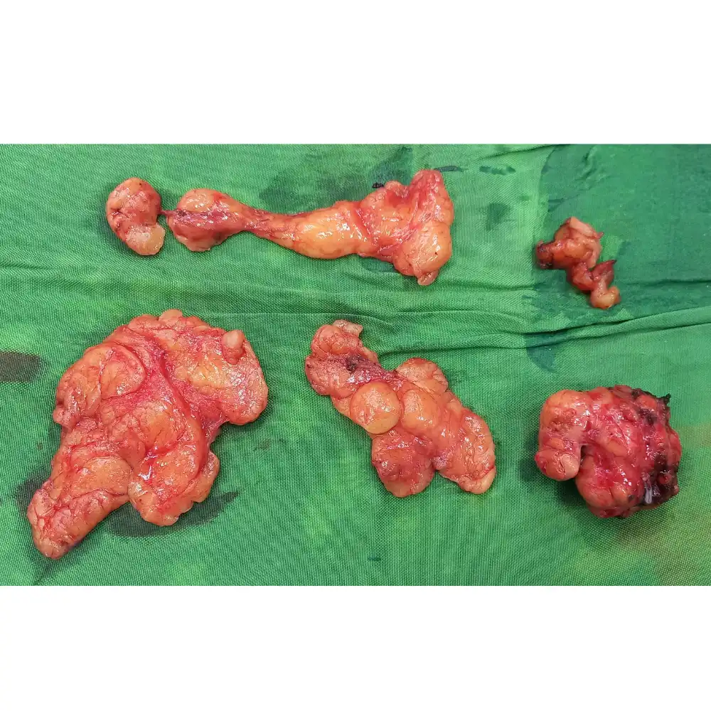
⦿ Right Radical Nephrectomy And Lymph Node Dissection
• Procedure: A radical right nephrectomy with adrenalectomy and para-aortic lymph node dissection was successfully executed.
• Complex Anatomy: The massive twenty-centimeter tumor was delicately separated from the compressed inferior vena cava.
• Patient Safety: Excellent hemostasis was maintained throughout the complex resection, minimizing blood loss to four hundred milliliters.
• Cancer Clearance: Pathological evaluation confirmed all surgical margins were completely negative for invasive carcinoma.
⦿ Successful Recovery And Complete Surgical Excision
The patient experienced an entirely uneventful post-operative recovery, rapidly regaining mobility and remaining completely hemodynamically stable. Final histopathology revealed an aggressive high-grade clear cell carcinoma with completely negative surgical margins, successfully achieving crucial local disease control. He was discharged home comfortably with excellent pain management, successfully resuming an independent and fully functional lifestyle.
Navigating such a massive tumor compressing vital vascular structures required extreme meticulousness and advanced surgical planning. By achieving perfectly clear margins alongside extensive lymph node clearance, we provided this young patient with the strongest possible foundation for long-term survival.
A 62-year-old female patient presented with an asymptomatic left renal mass discovered during routine screening. Imaging revealed a complex 5.3-centimeter lesion invading the upper pole of the left kidney. Prompt surgical intervention was crucial to remove this malignant tumor while carefully preserving healthy kidney tissue.
⦿ Robotic Left Partial Nephrectomy
• Procedure: A precise robotic left partial nephrectomy was executed utilizing the Da Vinci Xi system.
• Complex Anatomy: The upper pole tumor was meticulously separated while navigating the delicate left renal hilum.
• Safety Profile: Exceptional hemostasis was maintained throughout the procedure, resulting in a minimal blood loss of 300ml.
• Cancer Clearance: Histopathology confirmed complete tumor removal with negative resection margins for localized clear cell carcinoma.
⦿ Cancer-Free With Preserved Kidney Function
The patient experienced an exceptional and uneventful postoperative recovery, becoming fully ambulatory very shortly after her surgery. Final histopathology results confirmed a localized pT1b clear cell renal cell carcinoma with completely negative surgical resection margins. She was safely discharged in a hemodynamically stable condition, successfully retaining excellent baseline renal function.
By leveraging the enhanced 3D visualization of the robotic platform, we successfully eradicated the malignancy while maximizing nephron preservation. This precise approach ensures the patient remains cancer-free while safeguarding her long-term renal function.
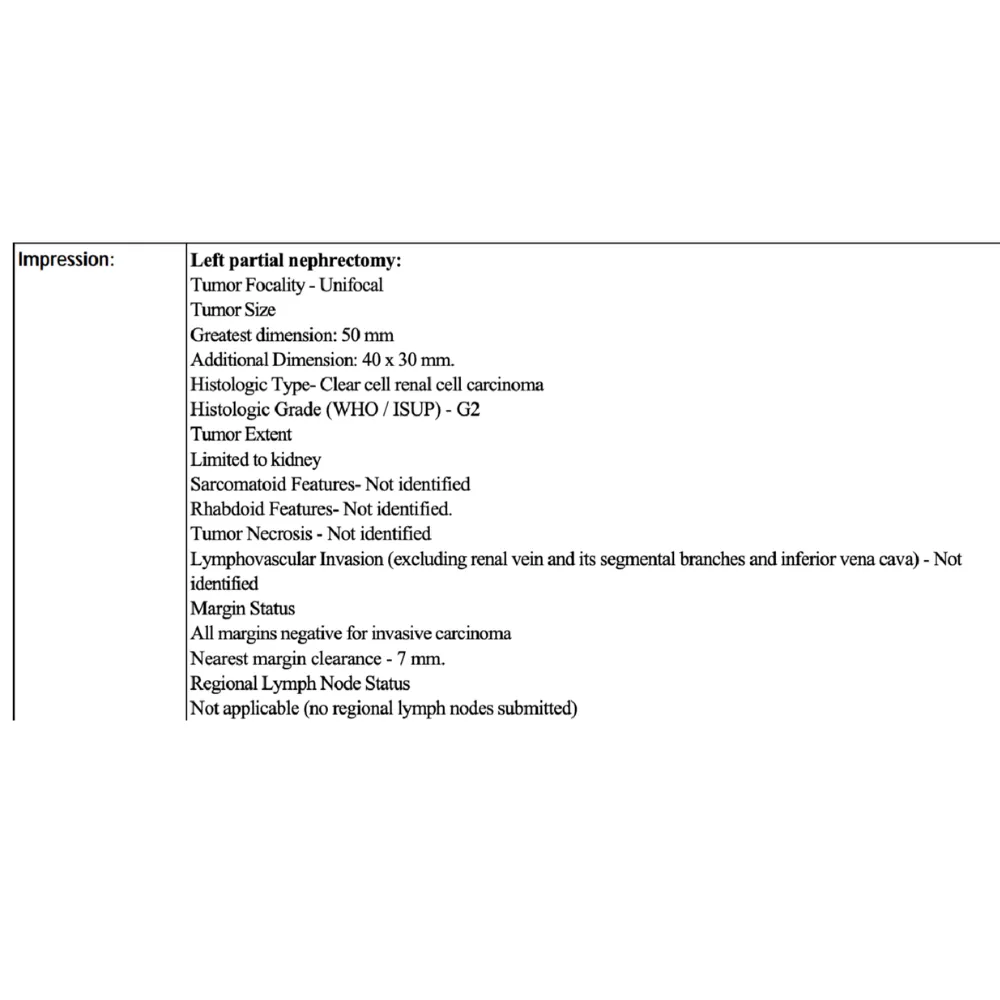
A 42-year-old female presented with a complex malignant left renal mass diagnosed as clear cell renal cell carcinoma. The 50 mm tumor posed a significant surgical challenge due to its substantial size. Preserving healthy kidney tissue while ensuring absolute cancer eradication was the primary clinical imperative.
⦿ Robotic-Assisted Left Partial Nephrectomy
• Procedure: Dr. Shah executed a robotic left partial nephrectomy with meticulous anatomical precision.
• Tumor Resection: The prominent 50 mm grade two clear cell carcinoma was completely excised.
• Patient Safety: Advanced robotic visualization ensured minimal blood loss and highly stable intraoperative hemodynamics.
• Cancer Clearance: Histopathology confirmed universally negative surgical margins with a safe clearance of 7 mm.
⦿ Excellent Recovery and Negative Margins
The patient experienced an exceptional postoperative recovery, remaining hemodynamically stable and mobilizing very shortly after surgery. Final histopathology confirmed a stage pT1b clear cell carcinoma with universally negative surgical margins. By retaining maximum healthy tissue, the patient successfully preserved excellent long-term renal function and achieved a stable discharge.
By leveraging advanced robotic precision, we successfully excised the complex 50 mm renal mass while thoroughly preserving the patient's healthy kidney. This nephron-sparing approach definitively treats the localized carcinoma and ensures an outstanding long-term quality of life.
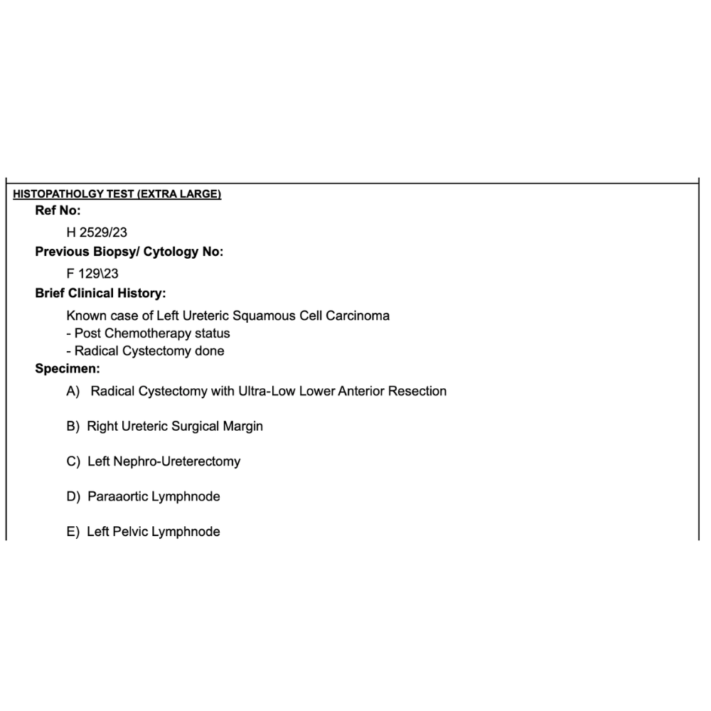
The patient, a 56-year-old male, presented with hematuria, weight loss, and an advanced left ureteric squamous cell carcinoma involving the urinary bladder and rectosigmoid junction. The aggressive tumor had formed a fistula and rendered the left kidney completely non-functional, requiring complex surgical intervention after extensive chemotherapy.
⦿ Left Nephrectomy with Exenteration
• Procedure Details: A meticulous left nephrectomy and radical exenteration were successfully executed under general anesthesia.
• Lymph Node Dissection: Extensive para-aortic and pelvic lymph node dissections were meticulously performed to accurately stage the cancer.
• Surgical Safety: The complex resection was completed with excellent hemostasis and stable hemodynamics throughout the entire procedure.
• Cancer Clearance: Frozen sections and final pathology confirmed absolute tumor-free surgical margins across all extensively resected organs.
⦿ Complete Resection and Functional Recovery
Following the extensive surgery, the patient's postoperative recovery was uneventful, discharging hemodynamically stable and ambulatory. Final histopathology confirmed a moderately differentiated squamous cell carcinoma, but importantly, all comprehensive resection margins were completely free of tumor. He achieved a safe discharge with a newly functioning ileal conduit and loop ileostomy.
Navigating this massive, multi-organ ureteric tumor required extreme precision and comprehensive exenteration to achieve completely clear margins. By meticulously executing this complex nephrectomy and reconstruction, we provided the patient with the absolute best opportunity for long-term survival and recovery.
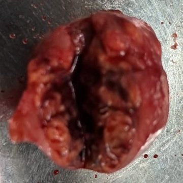
A 36-year-old male presented with severe abdominal pain and was subsequently diagnosed with a complex left renal mass. The tumor was located precariously near the spleen in the setting of concurrent acute pancreatitis, requiring a highly precise surgical approach to save the kidney while ensuring cancer removal.
⦿ Robotic Left Partial Nephrectomy
• Procedure: A robotic partial nephrectomy was flawlessly executed with a minimal thirty-minute warm ischemia time.
• Anatomical Complexity: The tumor was carefully detached from the adjacent enlarged spleen and adherent peripancreatic fat.
• Patient Safety: The advanced robotic approach ensured excellent hemostasis with a surprisingly low estimated blood loss.
• Cancer Clearance: Histopathology confirmed clear cell renal carcinoma with absolutely negative surgical margins for complete cure.
⦿ Excellent Oncological Clearance and Rapid Recovery
The patient experienced an exceptionally smooth postoperative recovery and was safely discharged home with completely stable hemodynamics. Final pathology definitively confirmed an early-stage clear cell renal cell carcinoma with absolutely negative surgical margins, indicating excellent cancer clearance. He quickly regained his normal ambulatory function and healed beautifully.
By utilizing the advanced robotic system, I was able to completely eradicate the localized renal tumor while safely sparing the patient's crucial kidney function. This precise nephron-sparing approach ultimately provided him with the absolute best oncological outcome and a rapid recovery.
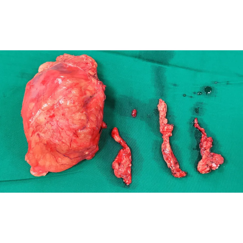
A 54-year-old female presented with hematuria and a large, multifocal right renal mass. Imaging revealed a highly complex 7-centimeter tumor with severe right renal vein thrombosis extending directly toward the inferior vena cava. This aggressive vascular involvement required immediate, highly specialized surgical intervention.
⦿ Right Radical Nephrectomy with IVC Thrombus Resection
• Procedure: The surgical team executed a flawless radical nephrectomy paired with delicate venous thrombus extraction.
• Complex Anatomy: Five para-aortic lymph nodes were completely dissected and proved entirely negative for tumor metastasis.
• Surgical Safety: The complex resection was completed with minimal blood loss totaling only one hundred milliliters.
• Cancer Clearance: The ureteral and arterial margins were preserved completely free of any invasive carcinoma cells.
⦿ Uneventful Recovery and Favorable Pathology
The patient experienced a remarkably smooth postoperative recovery and was safely ambulatory shortly after the complex procedure. Final pathology confirmed a completely resected clear cell renal cell carcinoma with negative regional lymph nodes and clean arterial margins. She was discharged in stable hemodynamic condition with an excellent foundation for long-term oncological monitoring.
Tackling an aggressive renal tumor with direct vena cava extension requires absolute surgical precision and meticulous anatomical control. By successfully extracting the vascular thrombus alongside the primary malignancy, we have given this patient the strongest possible chance for a definitive cure and enduring survival.
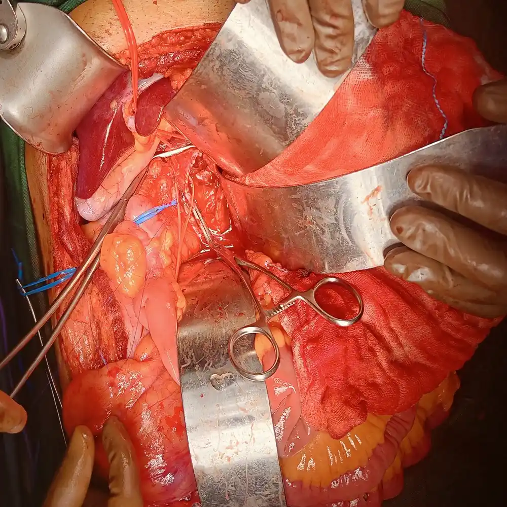
A 49-year-old male presented with a massive, non-functioning left renal mass measuring over fourteen centimeters. The cancer had aggressively invaded the left renal vein, forming a perilous thrombus extending directly into the inferior vena cava. This highly complex anatomy required immediate, meticulous surgical intervention.
⦿ Left Radical Nephrectomy with IVC Thrombus Resection
• Procedure: A left radical nephrectomy with IVC thrombus resection was flawlessly executed.
• Complex Anatomy: Eight para-aortic lymph nodes were carefully dissected to ensure comprehensive disease evaluation.
• Patient Safety: Meticulous vascular control maintained excellent patient safety with efficiently managed intraoperative blood loss.
• Cancer Clearance: Complete tumor removal was achieved, yielding definitively negative margins across all critical surgical boundaries.
⦿ Cancer-Free Margins and Excellent Clinical Recovery
Following the complex operation, the patient experienced a remarkably smooth and uneventful recovery. He was ambulatory shortly after surgery and discharged with completely stable hemodynamics. Final pathology confirmed clear cell renal cell carcinoma with zero lymph node spread and completely negative surgical margins. This comprehensive resection effectively eliminated the cancer while preserving critical vascular function.
Tackling a massive renal tumor with severe IVC involvement requires extreme surgical precision and careful vascular management. By meticulously isolating and removing the thrombus, we safely secured completely negative margins, giving this patient the absolute best chance at long-term survival.
A 50-year-old male presented with a one-month history of left flank pain and an incidental finding of a complex renal mass. Imaging revealed a 3.5-centimeter solid cystic lesion located in the interpolar region of the left kidney, resting precariously close to the delicate hilum and ureter.
⦿ Robotic Left Radical Nephrectomy
• Procedure: A five-port robotic transperitoneal approach was utilized for optimal anatomical visualization and maneuverability.
• Anatomical Challenge: The tumor's dangerous proximity to the renal hilum required extremely careful and controlled dissection.
• Blood Loss: The precise robotic surgical approach resulted in a minimal blood loss of just fifty milliliters.
• Cancer Clearance: Final pathology confirmed a completely negative margin status for the localized clear cell carcinoma.
⦿ Complete Eradication and Rapid Recovery
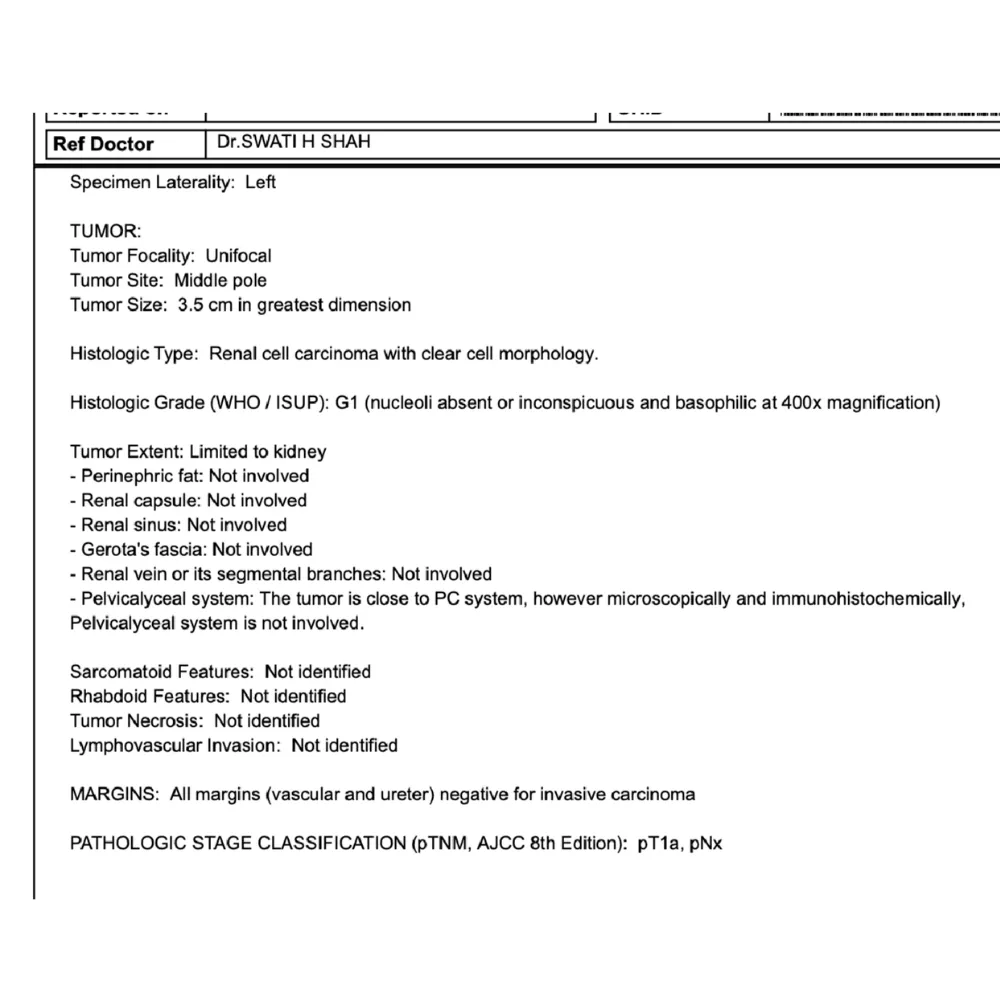
The patient experienced a remarkably smooth and uneventful postoperative recovery period with completely stable hemodynamics. Final histopathology revealed a well-differentiated, grade one clear cell renal carcinoma with entirely tumor-free vascular and ureteric margins. He regained full functional independence rapidly and was safely discharged just three days after the complex surgical intervention.
Managing tumors near the hilum demands absolute surgical precision to avoid catastrophic vascular injury. The robotic platform allowed us to safely navigate this challenging anatomy, securing a complete oncological cure while ensuring a fast, minimally invasive recovery for our patient.
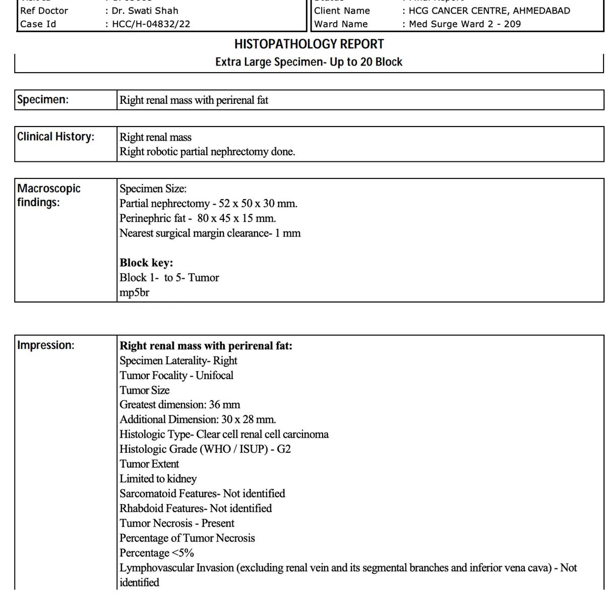
A fifty-eight-year-old female patient presented with a highly concerning right renal mass requiring urgent intervention. The malignant tumor measured nearly four centimeters in greatest dimension, demonstrating elements of tumor necrosis. The challenging anatomical location necessitated advanced surgical techniques to safely preserve the healthy functional kidney tissue.
⦿ Robotic Right Partial Nephrectomy
• Procedure: A robotic right partial nephrectomy was flawlessly executed to safely remove the unifocal tumor.
• Tumor Characteristics: The targeted right renal mass measured exactly thirty-six millimeters in its greatest dimension.
• Renal Preservation: The minimally invasive robotic approach minimized systemic blood loss while saving critical kidney tissue.
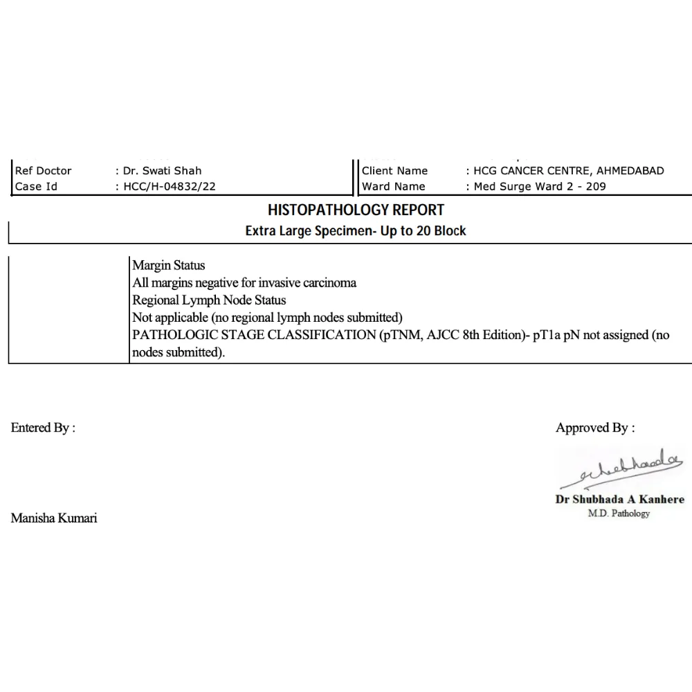
• Cancer Clearance: The final histopathology confirmed complete tumor removal with entirely negative surgical invasive margins.
⦿ Complete Surgical Cure and Renal Preservation
The patient recovered remarkably well, achieving complete hemodynamic stability and early ambulation shortly after the minimally invasive operation. Final histopathology definitively confirmed a stage pT1a clear cell renal cell carcinoma featuring entirely negative invasive surgical margins. The patient was safely discharged with successfully preserved renal function and excellent prospects for long-term survival.
By utilizing the advanced precision of the robotic system, we successfully eradicated the localized carcinoma while carefully sparing her functional kidney. This targeted organ-preservation strategy ensures excellent oncological control and provides the absolute best opportunity for a healthy, long-term recovery.
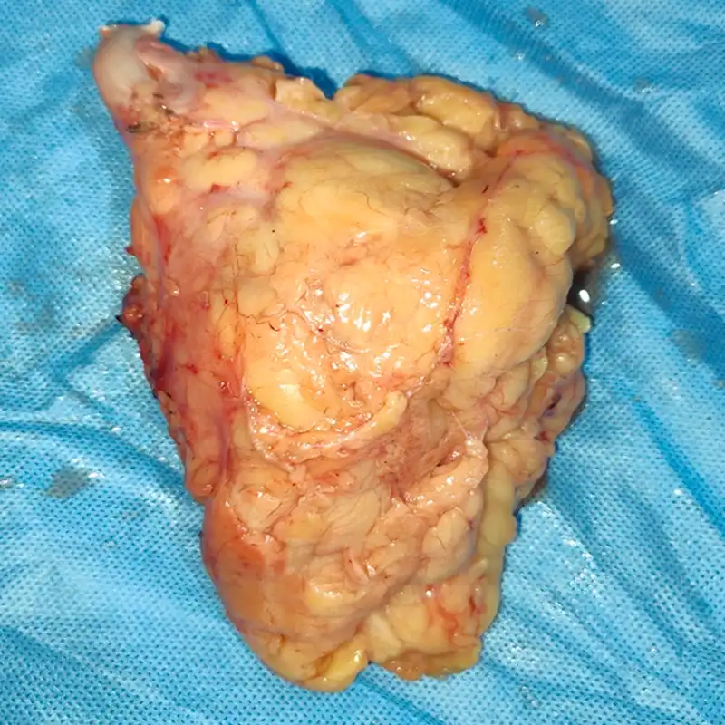
A 44-year-old female with a significant history of major pelvic cancer surgery presented with a new 9 cm mass in the left kidney. The surgical challenge was exceptionally high due to altered anatomy from prior bowel reconstruction and dense adhesions, requiring a specialized “redo” oncologic approach to isolate the renal vasculature safely.
⦿ Radical Nephrectomy with Retroperitoneal Lymphadenectomy
• Procedure: Dr. Shah executed a radical nephrectomy and para-aortic node dissection with ileal conduit revision.
• Complex Anatomy: Extensive adhesiolysis was required to navigate a 9 x 7 x 4 cm tumor in the mid-lower pole.
• Safety: Precision vascular control was maintained throughout the dissection, resulting in a controlled blood loss of 300 ml.
• Cancer Clearance: The surgical team achieved completely negative margins, ensuring no residual adenocarcinoma remained in the Gerota’s fascia.
⦿ Successful Tumor Removal and Functional Stability
The patient experienced a smooth postoperative recovery despite the high complexity of the redo surgery, remaining hemodynamically stable with well-managed pain. Final pathology confirmed a moderately differentiated adenocarcinoma confined to the kidney, with zero of the six examined lymph nodes showing metastasis. She resumed a normal diet gradually and was discharged in stable condition with an excellent long-term prognosis.
Redo oncologic surgeries in patients with prior exenterations require meticulous planning and advanced surgical precision. By successfully navigating dense adhesions and revised urinary pathways, we achieved complete tumor clearance, proving that even highly complex recurrent or secondary tumors can be managed effectively with specialized care.
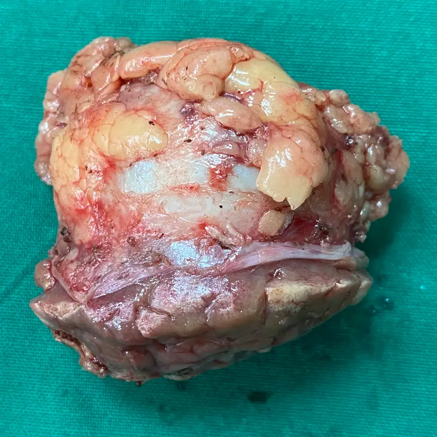
A 23-year-old male presented with an incidentally detected 4.5 cm right renal mass found during routine ultrasound imaging. The diagnosis of pT1b renal cell carcinoma in such a young patient demanded an urgent, nephron-sparing approach to ensure lifelong kidney function.
⦿ Robotic-Assisted Right Partial Nephrectomy
• Procedure: Executed a precise robotic partial nephrectomy with concurrent adhesiolysis and reconstructive pyeloplasty.
• Anatomical Challenge: Successfully isolated a single renal artery and vein to facilitate controlled vascular clamping.
• Safety / Blood Loss: Maintained exceptional patient safety with a minimal blood loss of only 300 ml.
• Cancer Clearance: Achieved total oncological clearance with confirmed negative margins and no lymphovascular invasion.
⦿ Complete Oncological Clearance and Recovery
The patient experienced a smooth postoperative recovery, achieving early ambulation and maintaining hemodynamic stability throughout his hospital stay. Final histopathology confirmed a Grade 2 clear cell renal cell carcinoma with perinephric fat entirely free of tumor. He was discharged in stable condition with fully preserved renal function and excellent long-term prognosis.
In younger patients, our primary goal is achieving a total cure while protecting long-term renal health through nephron-sparing techniques. This robotic approach allowed us to navigate complex anatomy with extreme precision, ensuring the patient remains cancer-free without compromising his quality of life.
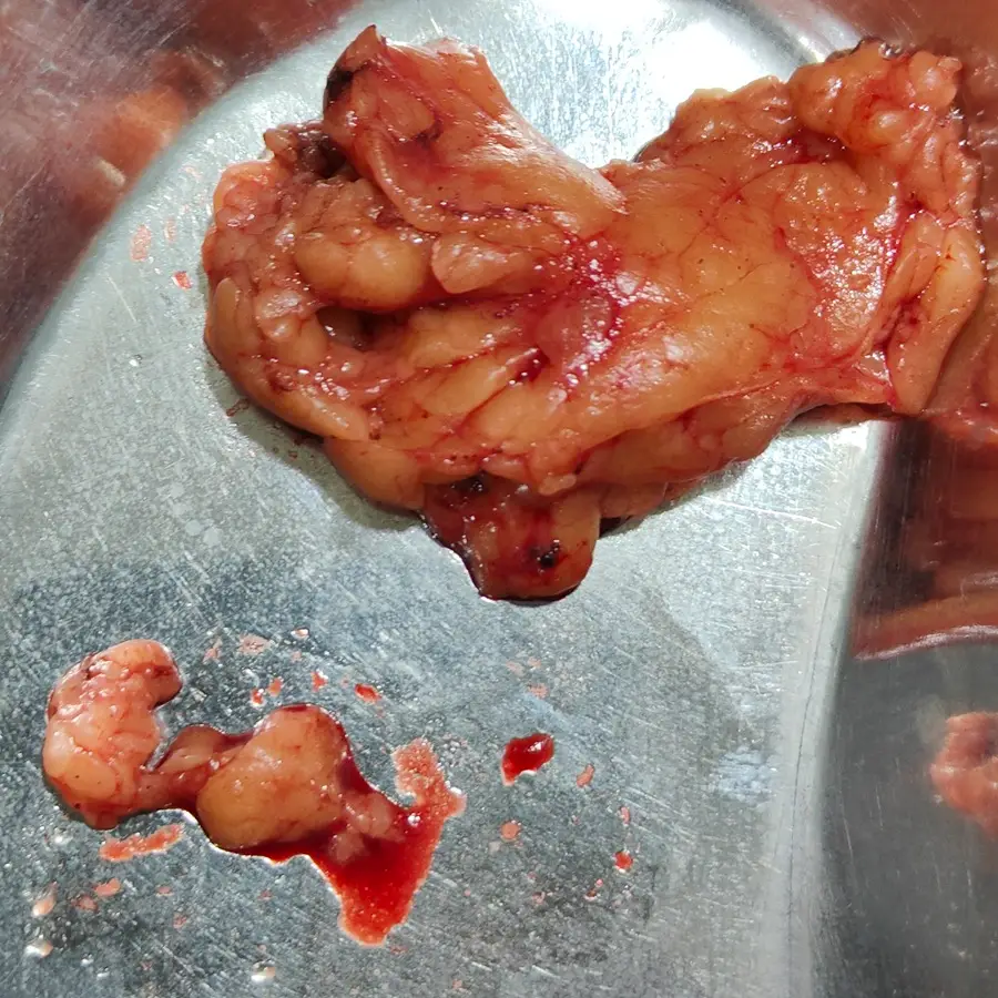
A 41-year-old male presented with flank pain and a 2.8 cm mass in the upper pole of his left kidney. Given the patient’s relatively young age and the localized pT1a stage, the clinical priority was to achieve complete oncological clearance while maximizing the preservation of healthy kidney tissue.
⦿ Robotic-Assisted Left Partial Nephrectomy
• Procedure: Executed a robotic-assisted partial nephrectomy combined with pyeloplasty and diagnostic laparoscopy.
• Anatomical Challenge: Targeted a well-defined 3 cm upper pole mass with precise vascular control.
• Safety / Blood Loss: Maintained exceptional patient safety, ensuring the patient remained hemodynamically stable throughout the procedure.
• Cancer Clearance: Achieved total tumor removal with confirmed negative surgical margins and no lymphovascular invasion.
⦿ Complete Oncological Cure and Organ Preservation
The patient’s recovery was excellent, characterized by early ambulation and effective pain management, leading to a stable discharge within just three days. Final histopathology confirmed a Grade 2 clear cell renal cell carcinoma confined to the kidney, with the perirenal fat entirely tumor-free. By preserving the majority of the left kidney, Dr. Shah ensured the patient maintains optimal long-term renal function and a superior quality of life.
In early-stage renal cancer, our goal is to deliver a definitive cure without sacrificing the patient's long-term kidney health. This robotic approach allows for the extreme precision needed to remove the tumor with clear margins while reconstructing the kidney for continued functional success.
Swasth Parivar: every week, practical advice on recovery, diet and warning signs. No advertising. Stop any time.
Your number is used for these updates and for the clinic to reach you. It is never sold or passed on. To stop, reply STOP on WhatsApp.