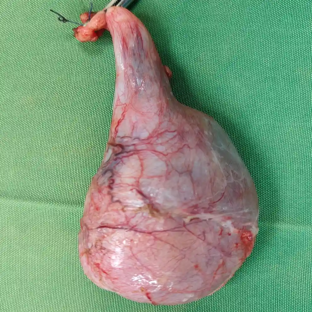
A 19-year-old male presented with rapid testicular swelling and abdominal discomfort persisting for nearly two weeks. Clinical evaluation revealed a 3.5 cm lesion and significantly elevated tumor markers, including AFP levels exceeding 1000, signaling an aggressive non-seminomatous germ cell tumor. The primary challenge was performing a swift intervention to halt the progression of this high-risk malignancy in a young patient.
⦿ High Inguinal Orchidectomy
• Procedure: A left high inguinal orchidectomy was performed to remove the primary germ cell tumor.
• Tumor Localization: The 3.5 cm firm mass was successfully resected while remaining strictly confined within the testis.
• Safety: The surgery was completed with minimal tissue trauma and zero intraoperative complications for the patient.
• Cancer Clearance: Radical excision was achieved with histologically confirmed negative margins along the vital spermatic cord.
⦿ Successful Resection and Rapid Recovery
The patient’s recovery was exceptionally smooth, allowing for independent ambulation and a successful discharge within only 24 hours of the procedure. Final pathology confirmed a pT2 stage with negative margins, providing a clear foundation for his multidisciplinary follow-up care. This timely surgical intervention successfully removed the primary source of the cancer, significantly optimizing his long-term curative potential.
For young patients with aggressive germ cell tumors, rapid surgical intervention via a high inguinal approach is the most critical step. We prioritized oncological safety to ensure complete primary tumor removal, which is essential for guiding effective long-term survival strategies.
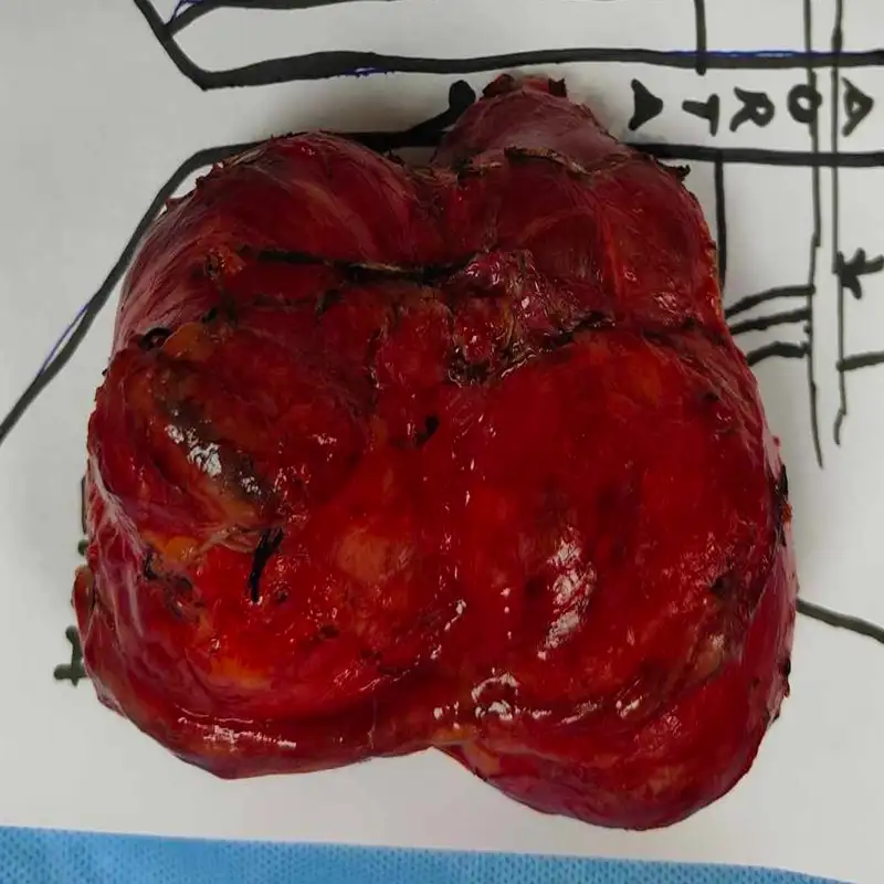
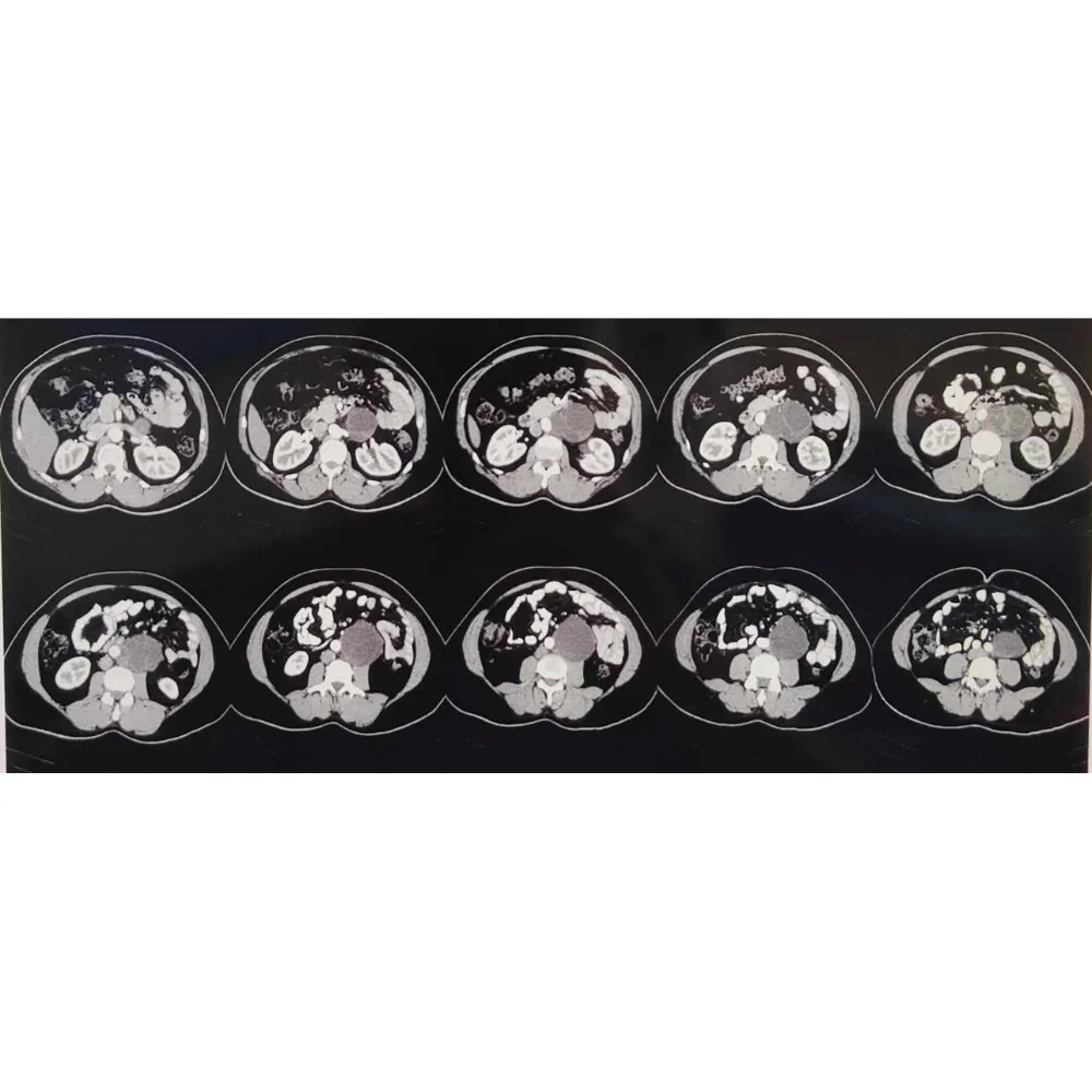
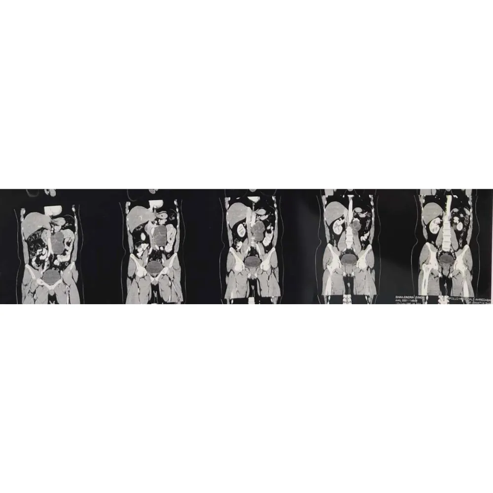
A 45-year-old male presented with persistent left flank pain following a previous high inguinal orchidectomy. Advanced imaging revealed a complex, 12.5 cm cystic mass in the para-aortic region, intricately abutting the renal vessels and aorta. The diagnosis of mature teratoma required urgent, high-precision surgical intervention to prevent further local invasion of vital abdominal structures.
⦿ Robotic Retroperitoneal Lymph Node Dissection (RPLND)
• Procedure: A comprehensive retroperitoneal lymph node dissection was executed with total mass excision.
• Complex Anatomy: The 14x10 cm mass was successfully separated from the aorta and renal vessels.
• Safety / Blood Loss: Exceptional surgical control resulted in a minimal blood loss of only 100 ml.
• Cancer Clearance: Complete resection was achieved, including the removal of fourteen reactive lymph nodes.
⦿ Complete Resection and Rapid Recovery
The patient’s postoperative recovery was uneventful, achieving hemodynamic stability and early mobilization by the fourth day. Final pathology confirmed a mature cystic teratoma with clear margins and no evidence of immature or malignant components. The patient was discharged in excellent condition with stable vitals and a transition to a soft, managed diet.
Treating large retroperitoneal teratomas requires absolute anatomical precision to protect the great vessels. By utilizing an advanced robotic approach, we achieved a complete oncological resection with minimal morbidity, ensuring the patient remains cancer-free while maintaining a high quality of life.
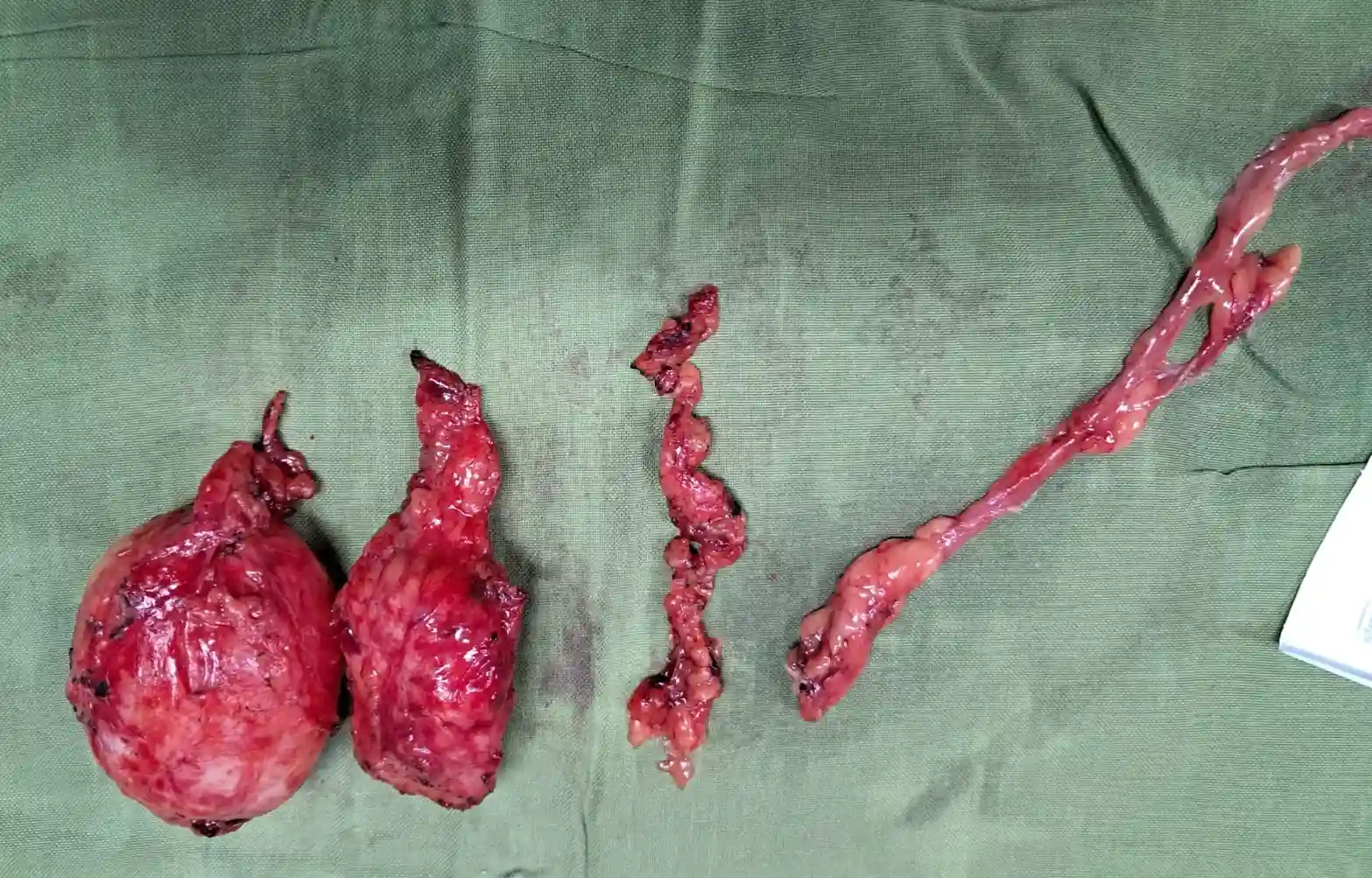
The patient is a 30-year-old male diagnosed with a high-grade metastatic germ cell tumor. Following intensive chemotherapy, imaging revealed a complex 4.5 cm calcified retroperitoneal mass and persistent pelvic lymphadenopathy. The case was highly complex due to the tumor’s proximity to major vascular structures and the necessity of complete surgical clearance for long-term survival.
⦿ Robotic Retroperitoneal Lymph Node Dissection (RPLND)
• Procedure: A full-template robotic retroperitoneal lymph node dissection was executed with absolute anatomical precision.
• Lymph Node Yield: Eleven regional lymph nodes were harvested, including the primary calcified retroperitoneal pelvic mass.
• Safety Profile: The robotic approach ensured minimal blood loss and preserved critical neural and vascular integrity.
• Cancer Clearance: Complete surgical resection was achieved, removing all cystic and calcified residual metastatic tumor foci.
⦿ Complete Surgical Remission
The patient demonstrated an excellent recovery, becoming hemodynamically stable and ambulatory within 24 hours post-surgery. Final pathology confirmed the removal of viable residual tumor elements within the lymph nodes, indicating a successful partial response to prior therapy followed by definitive surgical clearance. He was discharged with stable functional markers and a clear path toward long-term surveillance.
For young patients with residual germ cell tumors, robotic RPLND offers a superior alternative to traditional open surgery. By utilizing robotic precision, we achieved complete oncological clearance of calcified masses while significantly reducing recovery time and minimizing the risk of long-term functional morbidity.
A 28-year-old male presented with a significant residual retroperitoneal mass following intensive systemic chemotherapy for a non-seminomatous germ cell tumor. The primary challenge involved the tumor’s proximity to major vascular structures and the necessity of complete surgical resection to prevent potential malignant transformation or recurrence.
⦿ Nerve-Sparing Robotic RPLND
• Procedure: A full-template retroperitoneal lymph node dissection was executed using a minimally invasive robotic approach.
• Complex Anatomy: Precise dissection was required to isolate and preserve the sympathetic nerve fibers for functional maintenance.
• Safety & Blood Loss: Excellent hemodynamic stability was maintained throughout the procedure with minimal blood loss recorded.
• Cancer Clearance: Complete resection of the residual mass was achieved with clear surgical margins throughout.
⦿ Successful Recovery and Pathological Clearance
The patient recovered exceptionally well, mobilizing within twenty-four hours and demonstrating stable vital signs postoperatively. Final histopathology confirmed the successful removal of the mass with no evidence of viable malignancy in the remaining tissue. He was discharged shortly after, having regained full functional capacity and achieving a cancer-free status.
By utilizing the robotic platform for this RPLND, we achieved the extreme precision necessary to clear complex nodal disease while protecting vital nerves. This advanced approach ensures the most thorough oncological clearance while significantly accelerating the patient's return to their normal life.
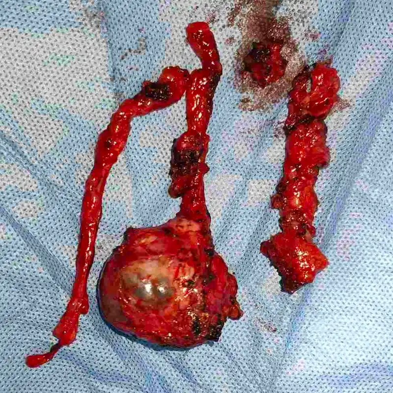
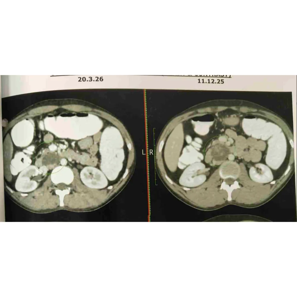
A 27-year-old male presented with residual retroperitoneal lymph node masses following multiple cycles of chemotherapy for recurrent non-seminomatous germ cell tumor of the right testis. The presence of a $3\times3$ cm solid-cystic mass adherent to the aorta required urgent surgical intervention to address potential chemo-resistant mature teratoma.
⦿ Robotic Retroperitoneal Lymph Node Dissection
• Procedure: A comprehensive full-template robotic retroperitoneal lymph node dissection was successfully executed.
• Complex Anatomy: Dr. Shah safely dissected the mass from the aorta and renal vasculature.
• Safety / Blood Loss: The surgery was completed with a minimal blood loss of only 200 ml.
• Cancer Clearance: Pathological analysis confirmed the removal of fourteen lymph nodes, including the mature teratoma.
⦿ Complete Resection and Rapid Recovery
The patient demonstrated excellent recovery, remaining hemodynamically stable and becoming ambulatory shortly after the procedure. Final histopathology confirmed a metastatic mature teratoma with post-therapy changes and negative margins in the surrounding iliac nodes and cord structures. He was discharged in stable condition with a clear plan for oncological follow-up.
For patients with residual masses following intensive chemotherapy, robotic RPLND offers the precision necessary to clear disease from major vessels. I focus on a full-template dissection to ensure we eliminate any teratoma components, providing the best path toward long-term survival.
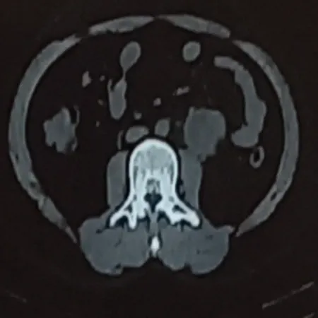
A 41-year-old male presented with a persistent $6\text{ cm}$ retroperitoneal mass and elevated CA 19-9 following a left orchidectomy. The complex lesion was strategically positioned at the L2-L3 level, significantly abutting the left ureter and the psoas muscle.
⦿ Robotic Retroperitoneal Lymph Node Dissection (RPLND)
• Procedure: Robotic-assisted retroperitoneal mass excision and systematic lymph node dissection were successfully executed.
• Complex Anatomy: The $6\text{ cm}$ mass encasing the inferior mesenteric artery was completely mobilized.
• Safety / Blood Loss: Exceptional surgical control resulted in a minimal blood loss of only 100ml.
• Cancer Clearance: Complete resection was achieved with pathological confirmation of a regressed germ cell tumor.
⦿ Seamless Recovery and Curative Pathology
Post-operative recovery was rapid and uneventful, with the patient achieving stable hemodynamics and early ambulation before discharge. Final histopathology confirmed a regressed germ cell tumor with mature teratomatous elements and no evidence of malignancy. The patient was discharged in excellent condition with fully preserved renal and gastrointestinal function.
By utilizing robotic precision, we successfully navigated the dense adhesions surrounding the major vessels and the ureter. This minimally invasive approach allowed for a complete oncological resection of the teratoma while ensuring the patient benefited from a significantly faster recovery.
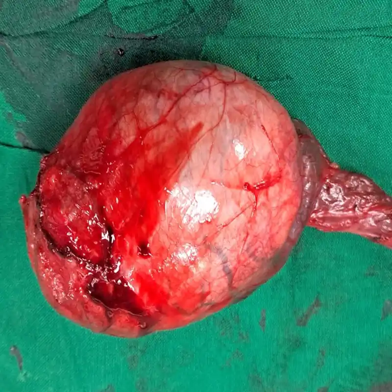
A 36-year-old male presented with a significant $7\times3$ cm hard, non-tender right testicular swelling persisting for one year. Diagnostic imaging revealed a large, complex lesion measuring $7.3\times6.3$ cm with elevated LDH and AFP markers. The case required urgent surgical intervention due to the tumor’s size and potential involvement of the scrotal wall.
⦿ Right High Inguinal Orchidectomy
• Procedure: A radical high inguinal orchidectomy was performed via an oblique incision.
• Complex Anatomy: The surgeon successfully isolated and ligated the spermatic cord at the deep ring.
• Safety / Blood Loss: The procedure was completed with minimal blood loss and no transfusions required.
• Cancer Clearance: Final pathology confirmed a pure classical seminoma with completely negative surgical margins.
⦿ Complete Oncological Remission
The patient experienced an excellent recovery and was ambulatory and hemodynamically stable shortly after the procedure. Histopathology confirmed a pT1b classical seminoma confined to the testis, with the spermatic cord and epididymis free of tumor. There was no evidence of lymphovascular invasion, and the patient was discharged in stable condition with a clear follow-up plan.
In cases of large testicular masses, a high inguinal approach is vital for achieving curative results. By performing deep ligation of the cord, we ensure complete oncological clearance. This precise surgical intervention effectively mitigated the risk of spread and secured an optimal functional outcome.
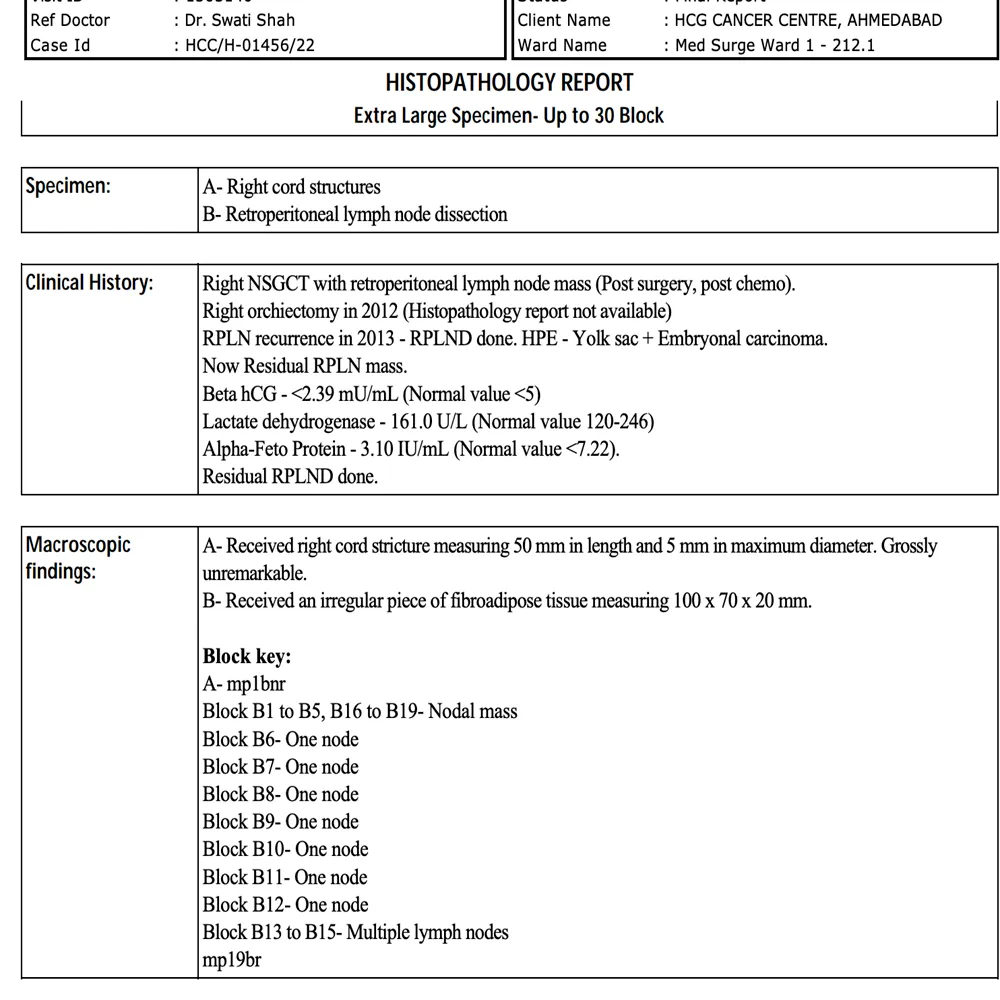
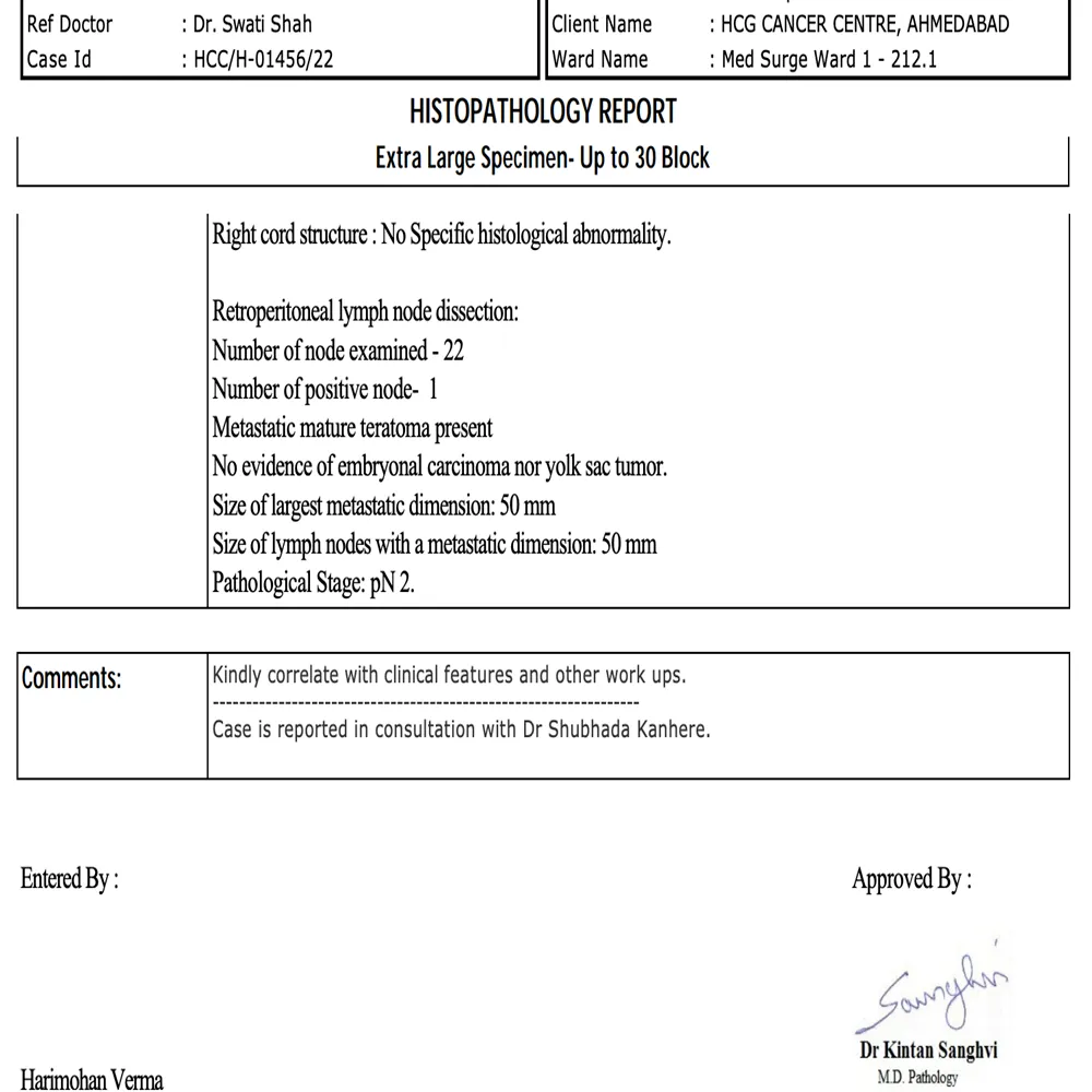
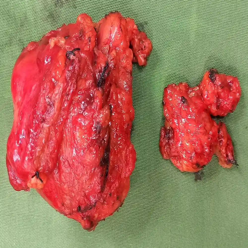
A 32-year-old male presented with a complex recurrence of Non-Seminomatous Germ Cell Tumor (NSGCT) following a prior orchiectomy and chemotherapy. The presence of a significant residual retroperitoneal mass required urgent, high-precision surgical intervention to address the chemo-resistant disease and prevent further local progression.
⦿ Robotic Residual RPLND
• Procedure: Robotic-assisted residual retroperitoneal lymph node dissection was executed with extreme precision.
• Nodal Assessment: A total of 22 lymph nodes were meticulously retrieved and examined.
• Safety Profile: The robotic platform facilitated minimal blood loss and enhanced patient safety.
• Cancer Clearance: Complete excision was achieved, successfully removing a 50mm metastatic mature teratoma.
⦿ Successful Clearance and Rapid Recovery
Post-operatively, the patient remained hemodynamically stable and was mobilized shortly after the procedure. Final histopathology confirmed the presence of a mature teratoma within the nodes, with no evidence of active embryonal carcinoma. The patient recovered exceptionally well and was discharged with stable renal function and excellent surgical outcomes.
By utilizing robotic precision, we successfully resected a complex, chemo-resistant residual mass in a difficult anatomical location. This advanced approach allowed for complete oncological clearance while ensuring the patient benefited from a significantly faster recovery and reduced morbidity.
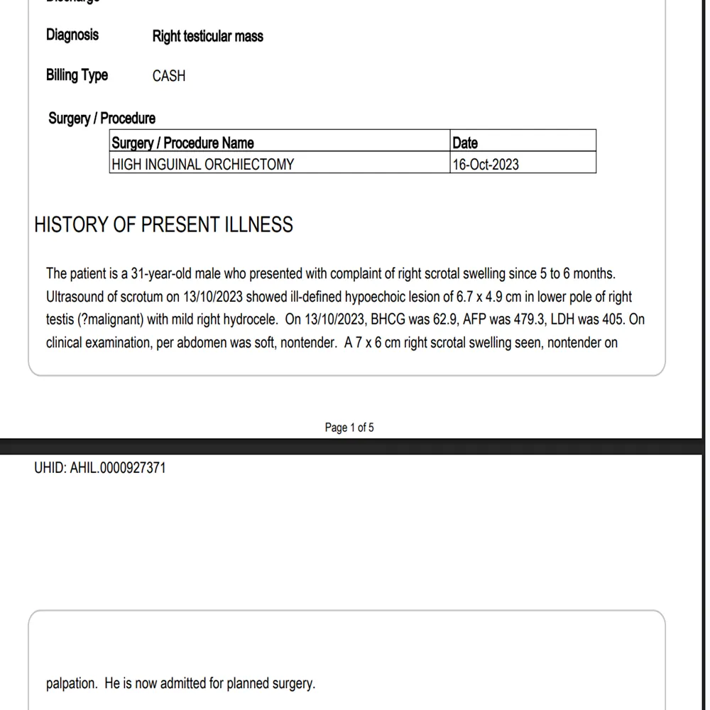
A 31-year-old male presented with a significant right scrotal swelling persisting for six months. Clinical evaluations and imaging confirmed a $6.7\times4.9$ cm hypoechoic mass with significantly elevated tumor markers, including an AFP of 479.3. The size and aggressive biochemical profile necessitated urgent, specialized oncological intervention.
⦿ Right High Inguinal Orchidectomy
• Procedure: A radical high inguinal orchidectomy was executed via a right inguinal incision.
• Vascular Control: The spermatic cord and testicular artery were ligated at the internal ring.
• Anatomical Handling: The testis was carefully mobilized from the scrotal sac by releasing the gubernaculum.
• Cancer Clearance: All surgical margins were confirmed negative for tumor involvement upon histopathological review.
⦿ Complete Resection and Rapid Recovery
Following the procedure, the patient’s postoperative recovery was entirely uneventful, allowing for a discharge within 24 hours. Pathology confirmed a Stage pT2 mixed germ cell tumor comprising 85% yolk sac tumor and 15% teratoma. The patient remained hemodynamically stable with a clean surgical wound and well-controlled pain levels at discharge.
In treating aggressive mixed germ cell tumors, a high inguinal approach is vital to ensure complete oncological clearance while preserving the integrity of the lymphatic pathways. By achieving negative margins and early vascular ligation, we provided this young patient with an optimal foundation for long-term survival.
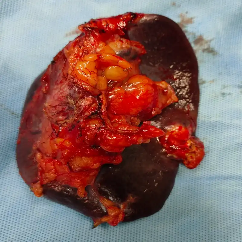
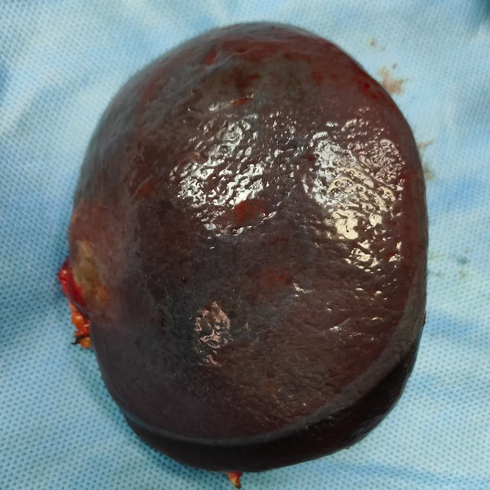
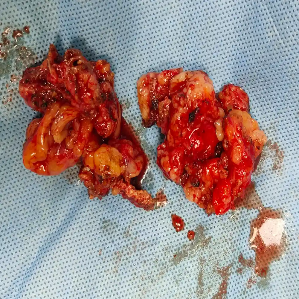
A 32-year-old male with a history of right testicular seminoma presented with residual retroperitoneal lymphadenopathy and a new splenic lesion following chemotherapy and radiotherapy. The clinical challenge involved navigating multiple enlarged nodes adherent to the aorta and IVC, alongside a 3 cm splenic mass involving the pancreatic tail, requiring a high-risk salvage resection.
⦿ Robotic RPLND, Splenectomy, and Pancreatectomy
• Procedure: Dr. Shah executed a complex robotic RPLND combined with a distal pancreatectomy and splenectomy.
• Complex Anatomy: The surgeon safely dissected nodal masses adherent to the aorta and inferior vena cava.
• Safety: Robotic precision allowed for a controlled blood loss of 250 ml during the multi-organ resection.
• Cancer Clearance: The procedure achieved a complete macroscopic clearance of the 3 x 2 cm splenic and pancreatic lesions.
⦿ Complete Pathological Response and Stable Recovery
The patient experienced a smooth postoperative recovery, achieving early mobilization and stable vital signs before being discharged within one week. Final histopathology confirmed a complete pathological response, with zero of the ten examined nodes containing viable tumor cells and the splenic lesion showing therapy-related necrosis. This exceptional oncologic result indicates a full disease clearance and provides a highly favorable long-term prognosis.
In cases of recurrent germ cell tumors, aggressive salvage surgery is often the final step toward a total cure. By performing a robotic RPLND and multi-organ resection, we successfully removed all residual masses, ultimately confirming that our combined treatment approach achieved a complete pathological response for this young patient.
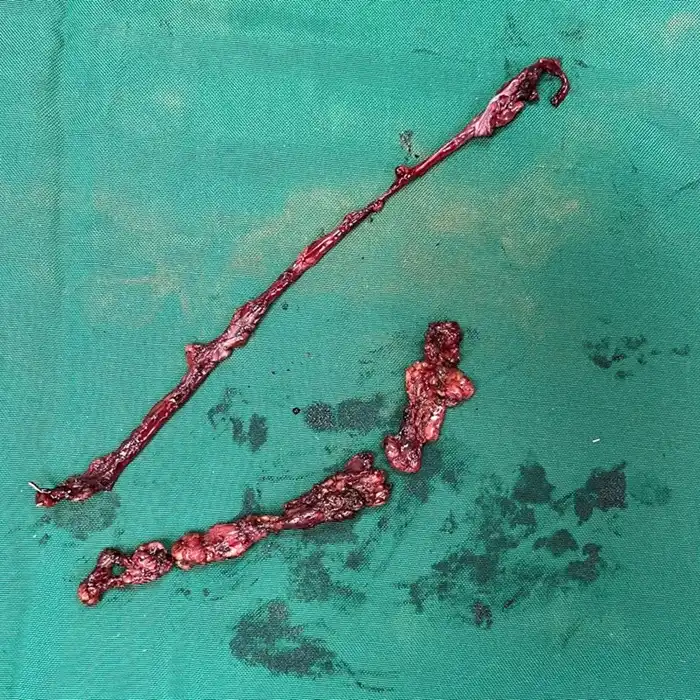
A 15-year-old male presented with a 5 cm left paratesticular mass, later confirmed as embryonal rhabdomyosarcoma. Following an initial inguinal orchidectomy, the primary clinical challenge involved performing an exhaustive retroperitoneal nodal staging to detect microscopic spread, which is essential for determining the intensity of subsequent pediatric chemotherapy protocols.
⦿ Robotic Nerve-Sparing RPLND and Chemoport Insertion
• Procedure: Dr. Shah executed a robotic nerve-sparing RPLND and left iliac lymph node dissection.
• Anatomy: The surgeon meticulously preserved the sympathetic nerve chains to maintain the teenager's future quality of life.
• Safety: The minimally invasive robotic approach allowed for a safe, bloodless dissection of the retroperitoneal space.
• Cancer Clearance: Fifteen total lymph nodes were harvested from the target zones to ensure accurate oncologic staging.
⦿ Pathological Node-Negative Status and Rapid Recovery
The patient experienced a smooth postoperative recovery, achieving early mobilization and stable vitals before a prompt discharge. Final histopathology confirmed that all fifteen harvested nodes were reactive and free of cancer, establishing a highly favorable prognosis for this Stage I, Group I malignancy. He transitioned to his planned chemotherapy regimen with a functional chemoport and excellent surgical healing of his minimally invasive port sites.
In pediatric oncology, achieving a cure must go hand-in-hand with preserving future function. By utilizing robotic nerve-sparing techniques for this RPLND, we provided the precise staging necessary to guide his chemotherapy while successfully protecting his long-term physical well-being.
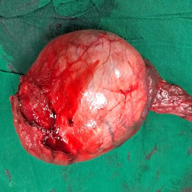
A 36-year-old male presented with a significant $7\times3$ cm hard, non-tender right testicular swelling persisting for one year. Diagnostic imaging revealed a large, complex lesion measuring $7.3\times6.3$ cm with elevated LDH and AFP markers. The case required urgent surgical intervention due to the tumor’s size and potential involvement of the scrotal wall.
⦿ Right High Inguinal Orchidectomy
• Procedure: A radical high inguinal orchidectomy was performed via an oblique incision.
• Complex Anatomy: The surgeon successfully isolated and ligated the spermatic cord at the deep ring.
• Safety / Blood Loss: The procedure was completed with minimal blood loss and no transfusions required.
• Cancer Clearance: Final pathology confirmed a pure classical seminoma with completely negative surgical margins.
⦿ Complete Oncological Remission
The patient experienced an excellent recovery and was ambulatory and hemodynamically stable shortly after the procedure. Histopathology confirmed a pT1b classical seminoma confined to the testis, with the spermatic cord and epididymis free of tumor. There was no evidence of lymphovascular invasion, and the patient was discharged in stable condition with a clear follow-up plan.
In cases of large testicular masses, a high inguinal approach is vital for achieving curative results. By performing deep ligation of the cord, we ensure complete oncological clearance. This precise surgical intervention effectively mitigated the risk of spread and secured an optimal functional outcome.
Swasth Parivar: twice a month, practical advice on recovery, diet and warning signs. No advertising. Stop any time.
Your number is used for these updates and for the clinic to reach you. It is never sold or passed on. To stop, reply STOP on WhatsApp.