
A recently managed case by Dr. Swati Shah, Uro Oncologist, involved a 71-year-old gentleman with PSA >100 ng/ml.
Pre-treatment evaluation showed:
⦿ PSMA expressive mass lesion involving nearly the entire prostate
⦿ Confluence of both seminal vesicles
⦿ Focally abutting the bladder neck
⦿ PSMA expressive metastatic pelvic, upper mesorectal and right obturator lymph nodes
⦿ No distant metastasis or skeletal metastasis.
⦿ Many patients believe pelvic lymph node involvement means surgery is no longer possible.
⦿ Current management has evolved.
In carefully selected patients with locally advanced prostate cancer (cN1) and disease confined to the pelvis, robotic radical prostatectomy with extended pelvic lymph node dissection can be an important part of multimodality treatment.
Proper evaluation by an experienced uro oncologist is essential before deciding between surgery, hormonal therapy, radiation or combined treatment.
A recently managed case by Dr. Swati Shah, Uro Oncologist, demonstrates that 71 years of age alone should not prevent curative prostate cancer surgery.
Following multidisciplinary evaluation, the patient underwent
Robotic Radical Prostatectomy + Bilateral Pelvic Lymph Node Dissection (PLND).
Robotic surgery offers several advantages in appropriately selected elderly patients:
In this case:
Treatment decisions should depend on fitness rather than age alone.
A recently managed prostate cancer case by Dr. Swati Shah, Uro Oncologist, highlights why complete cancer removal is the primary objective of surgery.
Pre-operative MRI showed:
⦿ Irregular T2 hypointense lesion
⦿ Patchy low ADC values
⦿ Heterogeneous enhancement
⦿ Loss of fat plane between lesion and bladder base
⦿ Involvement of the right seminal vesicle confluence
⦿ Enlarged right obturator/external iliac lymph nodes
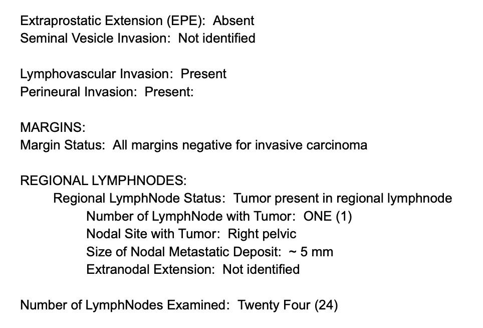
Despite these challenging findings, final histopathology after Radical Prostatectomy showed:
✅ All surgical margins negative for invasive carcinoma.
The specimen also demonstrated:
⦿ Acinar adenocarcinoma
⦿ Grade Group 4
⦿ Gleason Score 4+4=8
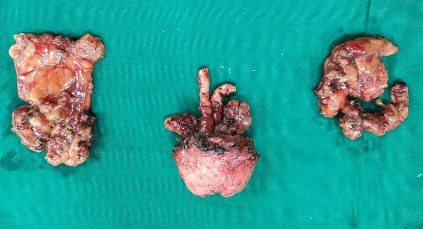
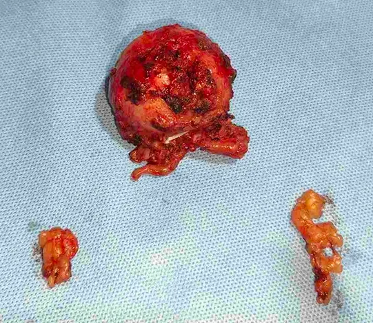
Robotic surgery provides excellent visualisation and meticulous dissection, helping achieve optimal oncological outcomes while preserving surrounding structures whenever oncologically safe.
A recently managed case by Dr. Swati Shah, Uro Oncologist, reminds us that PSA is only one part of prostate cancer assessment.
This patient initially presented with:
Following treatment and robotic surgery, final histopathology reported:
The Gleason Score (Grade Group) predicts how aggressive prostate cancer is and helps determine:
Patients searching for Gleason score, Grade Group 4 prostate cancer, PSA above 100, PI-RADS 5 lesion or PSMA PET-positive prostate cancer should understand that treatment decisions are based on the complete clinical picture—not PSA alone.
A recently managed prostate cancer case by Dr. Swati Shah, Uro Oncologist, highlights why pelvic lymph node dissection is performed during robotic radical prostatectomy in selected patients.
Pre-operative PSMA PET-CT demonstrated PSMA expressive metastatic pelvic, upper mesorectal and right obturator lymph nodes, while MRI also showed enlarged right obturator/external iliac lymph nodes.
Accordingly, the patient underwent:
Final histopathology showed:
Removing pelvic lymph nodes provides accurate staging, helps determine further treatment and may improve disease control in selected patients with node-positive prostate cancer.
A recently managed case by Dr. Swati Shah, Uro Oncologist, illustrates the role of neoadjuvant hormonal therapy in selected patients with locally advanced prostate cancer.
Initially:
The patient received one month of Abiraterone therapy, after which:
He subsequently underwent robotic radical prostatectomy with bilateral pelvic lymph node dissection.
Final histopathology also documented hormonal therapy effect present, confirming treatment response before surgery.
In carefully selected patients, systemic therapy followed by surgery can be an effective multimodality treatment strategy.
A recently managed prostate cancer case by Dr. Swati Shah, Uro Oncologist, demonstrates the advantages of robotic surgery in complex pelvic cancer.
The operation included:
The operation note documented:
Robotic prostate cancer surgery combines magnified three-dimensional vision with precise instrumentation, helping achieve meticulous cancer surgery while reducing blood loss and facilitating faster recovery.
A recently managed case by Dr. Swati Shah, Uro Oncologist, highlights recovery after robotic radical prostatectomy.
Following surgery:
In our routine practice, Foley catheter removal is generally performed after confirming satisfactory healing of the vesicourethral anastomosis, and urinary continence improves progressively with pelvic floor rehabilitation.
Many patients worry that recovery after prostate cancer surgery is prolonged. With modern robotic techniques, enhanced recovery protocols and experienced perioperative care, most patients are able to resume normal daily activities much earlier than expected.
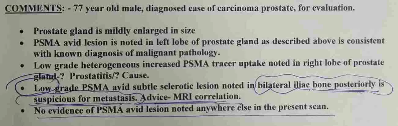
⦿ Recently managed was a 77-year-old man with carcinoma prostate, evaluated with 18F-PSMA PET/CT and MRI pelvis with contrast for treatment planning.
⦿ The PSMA PET/CT showed a PSMA-avid lesion in the left lobe of the prostate, measuring approximately 3.8 × 2.3 cm with SUV max 31.3. Low-grade heterogeneous PSMA uptake was also noted in the right lobe.
⦿ MRI provided further local assessment. It showed an irregular area of abnormal enhancement involving the left lateral half of the prostate, with involvement of both the peripheral and transition zones. The lesion measured approximately 3.3 × 3.2 × 2.2 cm.
⦿ Importantly, MRI showed no definite extra-prostatic extension, and both seminal vesicles were normal.
⦿ A few enlarged pelvic lymph nodes were also noted posterior to the external iliac arteries, the largest measuring approximately 2 × 0.9 cm.
⦿ Despite his age, the patient was considered for definitive surgical treatment after assessment of his overall health, tumour characteristics and staging.
⦿ He underwent robotic prostatectomy with bilateral pelvic lymph-node dissection.
⦿ Age by itself does not determine whether prostate cancer surgery is appropriate. A fit and functionally independent elderly patient may still be considered for curative treatment when the cancer characteristics and overall health are favourable.
⦿ Robotic prostatectomy can provide precise pelvic dissection with the potential advantages of reduced blood loss, faster recovery and earlier return to normal activity.
⦿ If you or a family member has been diagnosed with prostate cancer at an advanced age, treatment should be decided after considering the PSA, biopsy/Gleason Grade Group, MRI, PSMA PET-CT, overall health and life expectancy together.
Recently managed was a 77-year-old man with carcinoma prostate who underwent both 18F-PSMA PET/CT and MRI pelvis with contrast before definitive treatment.
The PSMA PET/CT showed a strongly PSMA-avid lesion in the left lobe of the prostate, approximately 3.8 × 2.3 cm, with a SUV max of 31.3.
However, PSMA PET-CT and MRI provide different types of information.
MRI demonstrated an irregular area of abnormal enhancement involving the left lateral half of the prostate, including:
MRI also demonstrated discrete enlarged lymph nodes posterior to the external iliac arteries, the largest measuring approximately 2 × 0.9 cm.
PSMA PET-CT helps identify PSMA-avid prostate cancer and possible disease elsewhere in the body.
MRI gives detailed anatomical information about the local extent of prostate cancer, including its relationship to the prostate capsule and surrounding structures.
This information is particularly important when considering robotic radical prostatectomy and planning pelvic lymph-node dissection.
In this patient, the imaging findings were integrated with the clinical and pathological information, and he underwent robotic prostatectomy with bilateral pelvic lymph-node dissection.
If your prostate cancer MRI mentions terms such as peripheral zone lesion, transition zone involvement, extra-prostatic extension, seminal vesicle involvement or pelvic lymph nodes, these findings need to be interpreted together with your biopsy and PSMA PET-CT.
A single scan should not be used in isolation to decide treatment.
Recently managed was a 77-year-old man with an established diagnosis of carcinoma prostate, who underwent MRI and 18F-PSMA PET/CT for staging and treatment planning.
This distinction is important because patients frequently ask:
“If my MRI or PSMA PET shows a suspicious lesion, do I still need a biopsy?”
For a new suspected prostate cancer, imaging usually does not replace tissue diagnosis.
A prostate biopsy provides important information including:
⦿ Confirmation of prostate cancer
⦿ Gleason score
⦿ Grade Group
⦿ Tumour grade and aggressiveness
⦿ Distribution of cancer within the sampled prostate tissue
⦿ These findings help determine whether the patient is suitable for active surveillance, surgery, radiotherapy or other treatment.
This patient was already diagnosed with carcinoma prostate.
The PSMA PET/CT demonstrated a PSMA-avid lesion in the left lobe of the prostate, with SUV max 31.3.
MRI subsequently demonstrated an irregular lesion involving the peripheral and transition zones, measuring approximately 3.3 × 3.2 × 2.2 cm.
MRI also assessed important local features:
No definite extra-prostatic extension was seen and both seminal vesicles were normal.
Pelvic lymph nodes were also assessed.
The imaging was therefore used for staging and treatment planning, followed by definitive surgical treatment with robotic prostatectomy and bilateral pelvic lymph-node dissection.
MRI can strongly suggest prostate cancer. PSMA PET-CT can help with staging.
But in a newly suspected case, histopathology from prostate biopsy provides the diagnosis and Grade Group/Gleason score that helps guide treatment.
If your report says “suspicious prostate lesion,” “PI-RADS lesion,” “PSMA-avid lesion” or “possible malignancy,” discuss whether biopsy is required before deciding on treatment.
⦿ MRI showed an irregular lesion involving the left lateral half of the prostate, with involvement of the peripheral and transition zones.
⦿ A few enlarged lymph nodes were seen posterior to the external iliac arteries, the largest approximately 2 × 0.9 cm, reported as possibly metastatic.
⦿ For many patients, the words “lymph node” or “possible metastasis” immediately create fear that surgery is no longer possible.
⦿ But the presence of suspicious pelvic lymph nodes does not, by itself, determine the entire treatment plan.
The decision requires assessment of:
In this case, MRI showed no definite extra-prostatic extension, and both seminal vesicles were normal.
After multidisciplinary assessment and staging, the patient underwent robotic prostatectomy with bilateral pelvic lymph-node dissection.
During prostate cancer surgery, removal of selected pelvic lymph nodes can provide important pathological staging information and help determine the patient’s subsequent treatment and follow-up strategy.
A suspicious pelvic lymph node on a scan does not automatically mean that prostate cancer surgery is impossible.
Every case needs individualized staging using the biopsy, Gleason Grade Group, MRI, PSMA PET-CT and clinical condition before deciding the best treatment.
For patients looking for prostate cancer surgery, robotic prostatectomy or treatment of prostate cancer with pelvic lymph nodes, specialist evaluation of the complete reports is essential.
⦿ Difficulty in passing urine is commonly caused by benign enlargement of the prostate, and TURP (transurethral resection of prostate) is often performed to relieve urinary obstruction. However, an important question before TURP is whether the patient could also have underlying prostate cancer.
⦿ In this case, a 70-year-old man presented with difficulty during micturition and underwent cystoscopy and TURP. Histopathology of the TURP specimen unexpectedly showed prostatic acinar adenocarcinoma, Gleason score 3+3=6, Grade Group 1. His PSA values were not markedly elevated.
⦿ This illustrates why PSA evaluation before prostate surgery is important. A low PSA does not completely exclude prostate cancer, and cancer may occasionally be discovered incidentally in TURP chips.
⦿ Once prostate cancer is identified in a TURP specimen, the patient needs appropriate assessment rather than assuming that TURP has treated the cancer. The extent of disease, PSA, imaging and the patient’s overall fitness need to be considered before deciding definitive treatment.
⦿ In this patient, further staging with Ga-68 PSMA PET-CT showed no obvious PSMA-avid lesion in the prostate, no significant PSMA-avid loco-regional adenopathy and no PSMA-avid distant metastasis. He was subsequently treated with robotic radical prostatectomy.
When prostate cancer is diagnosed, one of the important questions is whether the disease is confined to the prostate or has spread to pelvic lymph nodes or distant organs.
PSMA PET-CT is an advanced imaging investigation used for staging and treatment planning in appropriate patients with prostate cancer.
In this case, after prostatic acinar adenocarcinoma was identified following TURP, a whole-body Ga-68 PSMA PET-CT was performed. The report described:
These findings were important in deciding that there was no radiologically evident metastatic disease requiring treatment as metastatic prostate cancer.
A PSMA PET-CT, however, should not be interpreted in isolation. A negative scan does not completely exclude microscopic or very small-volume prostate cancer.
In this patient, the imaging findings were considered along with the TURP histopathology, PSA and clinical assessment before proceeding with robotic radical prostatectomy.
This is an example of how PSMA PET-CT can contribute to prostate cancer staging and surgical decision-making.
⦿ Prostate biopsy and TURP are performed for different purposes. TURP is primarily a procedure to relieve urinary obstruction, whereas a prostate biopsy is performed to obtain tissue specifically for diagnosing suspected prostate cancer.
⦿ This case demonstrates an important clinical scenario: the patient underwent TURP for difficulty in micturition, and the TURP specimen itself revealed prostatic acinar adenocarcinoma, Gleason score 3+3=6, Grade Group 1.
⦿ Therefore, if cancer is unexpectedly identified in the TURP specimen, the patient does not automatically need another biopsy simply to prove that cancer exists. The need for additional tissue sampling depends on what was found in the TURP specimen and whether further information is required for treatment planning.
⦿ The next step is appropriate prostate cancer risk assessment and staging, which may include PSA assessment, prostate imaging and, where indicated, PSMA PET-CT.
⦿ In this patient, the subsequent Ga-68 PSMA PET-CT did not demonstrate PSMA-avid loco-regional or distant metastatic disease. He was then evaluated for definitive treatment and underwent robotic radical prostatectomy.
⦿ Thus, an incidental prostate cancer diagnosis after TURP should lead to proper staging and treatment planning, rather than assuming that the TURP has completely treated the cancer.
Age alone should not decide whether an elderly patient can undergo prostate cancer surgery. Functional status, comorbidities, cancer characteristics, staging and expected benefit from treatment are all important.
This patient was 70 years old and had prostatic acinar adenocarcinoma diagnosed after TURP, with a Gleason score of 3+3=6, Grade Group 1. He had documented diabetes mellitus and hypothyroidism, but his functional evaluation showed that he was independent.
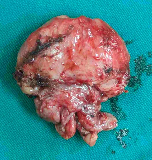
⦿ Recently managed was a 77-year-old man with carcinoma prostate, evaluated with 18F-PSMA PET/CT and MRI pelvis with contrast before definitive treatment.
⦿ Imaging showed a significant prostate lesion. The PSMA PET/CT demonstrated increased PSMA tracer uptake in the left lobe of the prostate, measuring approximately 3.8 × 2.3 cm with SUV max 31.3.
⦿ MRI demonstrated an irregular T2 hypointense lesion with patchy areas of low ADC values and heterogeneous patchy enhancement, involving the left lateral half of the prostate and both the peripheral and transition zones.
⦿ But imaging alone cannot tell us the complete biological behaviour of prostate cancer.
⦿ The Gleason score / Grade Group obtained from prostate biopsy helps determine how aggressive the prostate cancer is.
⦿ Two patients can have similar-looking prostate lesions on MRI but have very different tumour biology depending on their Gleason Grade Group.
⦿ Treatment decisions therefore need to consider: PSA + Gleason Grade Group + MRI + PSMA PET/CT + clinical condition
⦿ In this patient, MRI also assessed the local extent of disease and reported no definite extra-prostatic extension, with both seminal vesicles appearing normal.
⦿ Pelvic lymph nodes were also identified and considered in staging.
⦿ The patient was subsequently treated with robotic prostatectomy and bilateral pelvic lymph-node dissection.
If you have been diagnosed with prostate cancer, do not look at the MRI or PET report alone.
Your biopsy Gleason score/Grade Group is one of the most important pieces of information for deciding how aggressively the cancer needs to be treated.
⦿ The lesion involved both the peripheral and transition zones and measured approximately 3.3 × 3.2 × 2.2 cm.
⦿ The MRI specifically assessed whether the tumour had extended outside the prostate.
⦿ The report noted: “No extra-prostatic extension of the disease.”
⦿ Both seminal vesicles were also reported as normal.
When planning prostate cancer treatment, MRI is used not only to locate the tumour but also to assess its relationship with:
⦿ Prostate capsule
⦿ Surrounding tissues
⦿ Bladder base
⦿ Seminal vesicles
⦿ Neurovascular structures
⦿ Pelvic lymph nodes
A report showing no definite extra-prostatic extension provides important local staging information.
However, this does not mean that the cancer is automatically low-risk or completely confined microscopically. MRI findings must always be interpreted together with the PSA, biopsy Gleason Grade Group and PSMA PET/CT.
In this patient, PSMA PET/CT demonstrated a strongly PSMA-avid lesion with SUV max 31.3, while MRI provided detailed anatomical assessment.
After complete evaluation, the patient underwent robotic prostatectomy with bilateral pelvic lymph-node dissection.
When reading your prostate MRI, terms such as “extra-prostatic extension,” “seminal vesicle involvement,” “capsular involvement” and “pelvic lymph nodes” are important because they help determine the stage and treatment strategy.
⦿ Recently managed was a 77-year-old man with carcinoma prostate whose staging investigations showed suspicious pelvic lymph nodes.
⦿ MRI demonstrated discrete enlarged lymph nodes posterior to the external iliac arteries, with the largest measuring approximately 2 × 0.9 cm, described as possibly metastatic.
⦿ The PSMA PET/CT also played an important role in evaluating the prostate and looking for disease elsewhere.
⦿ When prostate cancer is being treated surgically, assessment of the pelvic lymph nodes can be an important part of accurate staging.
⦿ During the robotic operation, selected pelvic lymph nodes are removed and sent for histopathological examination.
⦿ This can determine whether prostate cancer has actually spread to the lymph nodes and provides more accurate pathological staging.
⦿ In this patient, robotic prostatectomy was performed along with bilateral pelvic lymph-node dissection.
The final pathology helps the treating team determine:
⦿ Whether lymph nodes contain cancer
⦿ The pathological stage
⦿ Whether additional treatment or closer surveillance may be required
⦿ The appropriate follow-up strategy
A suspicious lymph node on MRI does not automatically prove metastasis.
Imaging raises suspicion; histopathology provides confirmation.
If your prostate MRI or PSMA PET-CT mentions external iliac, obturator or pelvic lymph nodes, the significance depends on their size, morphology, tracer uptake and the overall cancer profile.
For patients with prostate cancer and suspicious pelvic lymph nodes, treatment should be planned after integrating biopsy, Gleason Grade Group, PSA, MRI and PSMA PET-CT findings.
⦿ Recently managed was a 77-year-old man with carcinoma prostate with a PSMA-avid primary prostate lesion and suspicious pelvic lymph nodes.
⦿ The MRI showed a 3.3 × 3.2 × 2.2 cm irregular lesion involving the left lateral half of the prostate, including the peripheral and transition zones.
⦿ The report also specifically documented no definite extra-prostatic extension and normal seminal vesicles.
⦿ When robotic prostatectomy is considered, the objective is not simply to remove the prostate.
⦿ The surgery requires precise dissection around important pelvic structures while achieving adequate cancer clearance.
Robotic surgery provides:
⦿ High-definition magnified vision
⦿ Precise instrument movements
⦿ Fine dissection in the confined pelvic space
⦿ Better visualization of important anatomical planes
⦿ Precise suturing for reconstruction
These advantages can be particularly useful when operating close to structures that are important for urinary continence and sexual function.
In this case, robotic prostatectomy with bilateral pelvic lymph-node dissection was performed after complete clinical and imaging assessment.
The priority in prostate cancer surgery remains complete cancer removal with an adequate oncological margin. Functional preservation is important, but it should never compromise cancer control.
The final assessment therefore depends on the histopathology of the prostate and lymph nodes, including the surgical margins and pathological stage.
For patients considering robotic prostatectomy for prostate cancer, the important question is not simply whether surgery is robotic.
The important questions are:
Is surgery appropriate for my stage of cancer? Can the tumour be removed safely? Can adequate cancer clearance be achieved? And how can urinary and sexual function be preserved without compromising cancer control?
⦿ A common concern after a patient is diagnosed with prostate cancer following TURP is whether previous prostate surgery makes a later radical prostatectomy difficult or impossible.
⦿ This 70-year-old man had undergone TURP on 14 March 2026 for difficulty in micturition. Histopathology showed prostatic acinar adenocarcinoma, Gleason score 3+3=6, Grade Group 1.
⦿ He subsequently underwent robotic radical prostatectomy.
⦿ The operative note specifically documented post-TURP status. During surgery, the bladder neck was reconstructed, a modified Rocco posterior reconstruction was performed and a vesicourethral anastomosis was completed. Bilateral neurovascular bundles were preserved.
⦿ Previous TURP can alter the normal anatomy around the bladder neck and prostate. Therefore, radical prostatectomy after TURP requires careful surgical planning and experience with the altered anatomy.
⦿ This case demonstrates that robotic radical prostatectomy can be performed after TURP in appropriately selected patients.
One of the most important goals of radical prostatectomy for prostate cancer is complete removal of the cancer with negative surgical margins.
In this 70-year-old patient, robotic radical prostatectomy was performed after prostate cancer was diagnosed following TURP.
The operation included careful dissection of the prostate, preservation of both neurovascular bundles, division of the urethra and completion of the prostatectomy. Bladder-neck reconstruction, posterior reconstruction and vesicourethral anastomosis were then performed.
The final histopathology was particularly reassuring:
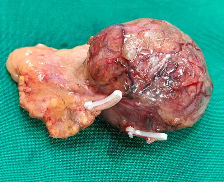
A negative surgical margin means that no tumour was identified at the cut edge of the removed specimen. It is an important pathological indicator after prostate cancer surgery and forms part of the overall assessment for future follow-up.
⦿ Surgery is only one part of prostate cancer treatment. The final histopathology report provides important information about what was found in the removed prostate and helps determine subsequent follow-up.
⦿ In this case, the prostatectomy specimen was examined extensively, including the apical and base margins, seminal vesicles, vas deferens and multiple sections of prostatic tissue.
⦿ Histologic type: Acinar adenocarcinoma was the known diagnosis, but no residual tumour was identified in the prostatectomy specimen, attributed to the post-TURP status.
⦿ No extraprostatic extension
⦿ No urinary bladder-neck invasion
⦿ No seminal-vesicle invasion
⦿ No lymphovascular invasion
⦿ No perineural invasion
⦿ No intraductal carcinoma
⦿ No cribriform glands
⦿ All assessed surgical margins negative for invasive carcinoma
This is why the prostatectomy specimen should not simply be reported as “prostate cancer removed.”
The pathology report answers several important questions about tumour extent, margins and pathological risk, which are essential for deciding the patient’s follow-up strategy.
In this patient, the final report was particularly favourable, with negative margins and no identified residual tumour in the prostatectomy specimen.
⦿ “How quickly can I recover after robotic prostatectomy?” is one of the most common questions patients ask before surgery.
⦿ This 70-year-old patient underwent robotic radical prostatectomy on 30 April 2026. The operative procedure included robotic prostate removal, bladder-neck reconstruction, posterior reconstruction and vesicourethral anastomosis.
⦿ His postoperative recovery was uncomplicated in the documented hospital course.
⦿ He was haemodynamically stable
⦿ He was ambulatory
⦿ Blood sugar was controlled
⦿ Pain was controlled
⦿ Surgical wound was clean
⦿ He was tolerating a normal diet
⦿ He was discharged on postoperative day 3
⦿ The urinary catheter remained in situ at discharge
The patient was advised routine physical activity, wound dressing after 3 days and oncology follow-up after 7 days.
Recovery after robotic prostatectomy is not measured only by the day of discharge. Urinary catheter management, wound healing, urinary continence, erectile function and PSA follow-up are all important parts of recovery.
This case shows that even a 70-year-old patient can have an early postoperative recovery after robotic radical prostatectomy when appropriately selected and managed.
⦿ Recently managed case by Dr Swati Shah – Robotic Uro-Oncology.
⦿ A 70-year-old patient was diagnosed with prostatic acinar adenocarcinoma, Gleason score 3+3=6, Grade Group 1, incidentally identified in the TURP specimen performed for difficulty in micturition.
⦿ A common concern is whether radical prostatectomy can be performed after TURP.
⦿ Previous TURP can alter the normal anatomy around the prostate and bladder neck, making subsequent surgery technically more demanding.
⦿ In this case, robotic radical prostatectomy was performed despite the previous TURP.
⦿ The operative findings specifically documented post-TURP status, with no evident extraprostatic spread, seminal vesicle invasion or lymphadenopathy.
⦿ Bilateral neurovascular bundle preservation
⦿ Bladder-neck reconstruction
⦿ Modified Rocco posterior reconstruction
⦿ Vesicourethral anastomosis
⦿ Pelvic lymph-node dissection
The robotic platform allows precise dissection and reconstruction in the confined pelvic space, which can be particularly useful when operating in altered anatomy after previous prostate surgery.
This case demonstrates that previous TURP does not automatically exclude robotic radical prostatectomy. Careful surgical planning and experience with post-TURP anatomy are important.
⦿ Recently managed case by Dr Swati Shah – Robotic Uro-Oncology.
⦿ The patient had prostatic acinar adenocarcinoma, Gleason 3+3=6, Grade Group 1, diagnosed following TURP.
⦿ During robotic radical prostatectomy, bilateral neurovascular bundle preservation was performed.
⦿ At the same time, the objective of prostate cancer surgery is not simply preservation of function—it is complete oncological removal of the tumour.
⦿ The operation included meticulous dissection of the prostate, bladder-neck reconstruction, posterior reconstruction and vesicourethral anastomosis.
⦿ No residual tumour identified in the prostatectomy specimen
⦿ No extraprostatic extension
⦿ No bladder-neck invasion
⦿ No seminal-vesicle invasion
⦿ No lymphovascular invasion
⦿ No perineural invasion
⦿ All surgical margins negative for invasive carcinoma
The apical, bladder-neck and circumferential margins were free of invasive carcinoma.
This highlights two important goals of robotic prostate cancer surgery:
Oncological control — complete tumour removal with negative margins and Functional preservation — careful preservation of the neurovascular bundles when oncologically appropriate.
Both have to be considered together when planning radical prostatectomy.
⦿ Recently managed case by Dr Swati Shah – Robotic Uro-Oncology.
The final pathology report after prostate cancer surgery provides much more information than simply stating whether cancer is present.
In this patient, prostate cancer had initially been diagnosed in the TURP specimen as prostatic acinar adenocarcinoma, Gleason score 3+3=6, Grade Group 1.
After robotic radical prostatectomy, the entire prostatectomy specimen was examined.
The final report documented: No residual tumour identified in the prostatectomy specimen, in the setting of previous TURP.
⦿ No extraprostatic extension
⦿ No urinary bladder-neck invasion
⦿ No seminal-vesicle invasion
⦿ No lymphovascular invasion
⦿ No perineural invasion
⦿ No intraductal carcinoma
⦿ No cribriform glands
⦿ Negative surgical margins
The vas deferens, seminal vesicles, apical margin and base margin were also free of invasive carcinoma.
This is why the final histopathology after radical prostatectomy is an essential part of prostate cancer management.
It helps establish the pathological status of the removed specimen and provides information that is important for subsequent PSA surveillance and follow-up planning.
Recently managed case by Dr Swati Shah – Robotic Uro-Oncology.
Recovery after robotic radical prostatectomy is an important concern for patients and families.
In this case, despite the patient’s age and medical history, the postoperative course was stable.
⦿ Haemodynamically stable
⦿ Ambulatory at discharge
⦿ Tolerating diet
⦿ Pain controlled
⦿ Surgical wound clean
⦿ Blood sugar controlled
He was discharged with the Foley catheter in situ, with follow-up planned.
The operative procedure involved robotic radical prostatectomy with bladder-neck reconstruction, posterior reconstruction and vesicourethral anastomosis.
Recovery after robotic prostatectomy is not limited to the hospital stay. Patients commonly want to know about:
walking after prostatectomy → catheter removal → wound recovery → urinary continence → PSA follow-up → return to routine activities.
Early mobilisation, appropriate pain control, catheter care and structured postoperative follow-up are important components of recovery.
This case demonstrates that robotic radical prostatectomy can be followed by early mobilisation and discharge even in an older patient when appropriately selected and managed.
Swasth Parivar: twice a month, practical advice on recovery, diet and warning signs. No advertising. Stop any time.
Your number is used for these updates and for the clinic to reach you. It is never sold or passed on. To stop, reply STOP on WhatsApp.