A recently managed germ cell tumour of the testis by Dr. Swati Shah, Uro Oncologist, helps explain one of the most common questions patients ask:
“Why has my cancer spread to the abdomen when the testis has already been removed?”
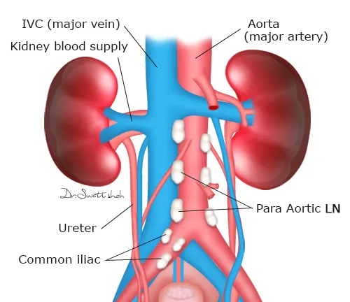
⦿ The answer lies in normal embryology. The testes develop high in the abdomen before descending into the scrotum during fetal life. However, their lymphatic drainage remains connected to the retroperitoneal para-aortic, interaortocaval, precaval and retrocaval lymph nodes, not the groin.
⦿ Therefore, when testicular cancer spreads, the retroperitoneal lymph nodes are usually the first site of metastasis. Enlargement of these nodes does not automatically mean widespread disease, and many patients can still achieve long-term cure with appropriate combinations of chemotherapy and surgery.
⦿ Understanding this predictable pattern of spread is essential for accurate staging, imaging interpretation and planning treatment, including Retroperitoneal Lymph Node Dissection (RPLND) when indicated.
A recently managed advanced testicular cancer case by Dr. Swati Shah, Uro Oncologist, demonstrated why Retroperitoneal Lymph Node Dissection (RPLND) is among the most technically demanding urologic cancer surgeries.
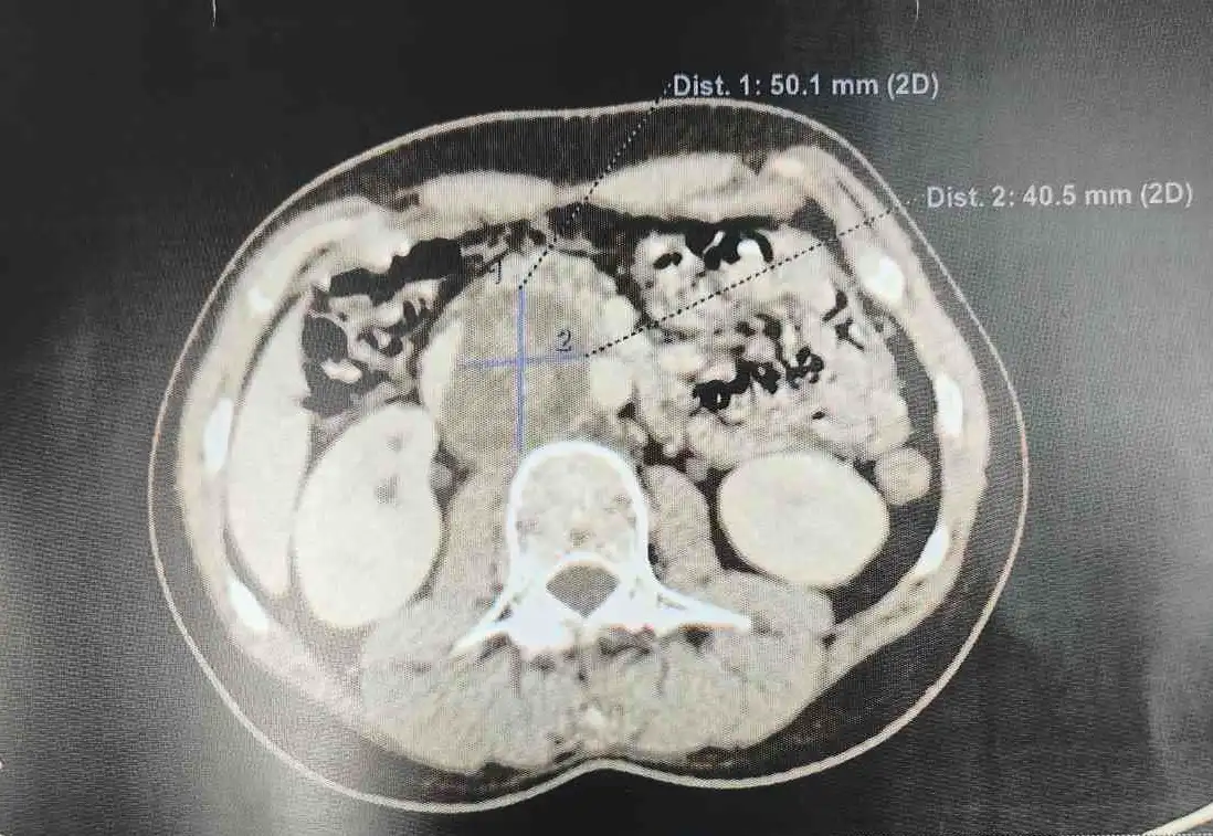
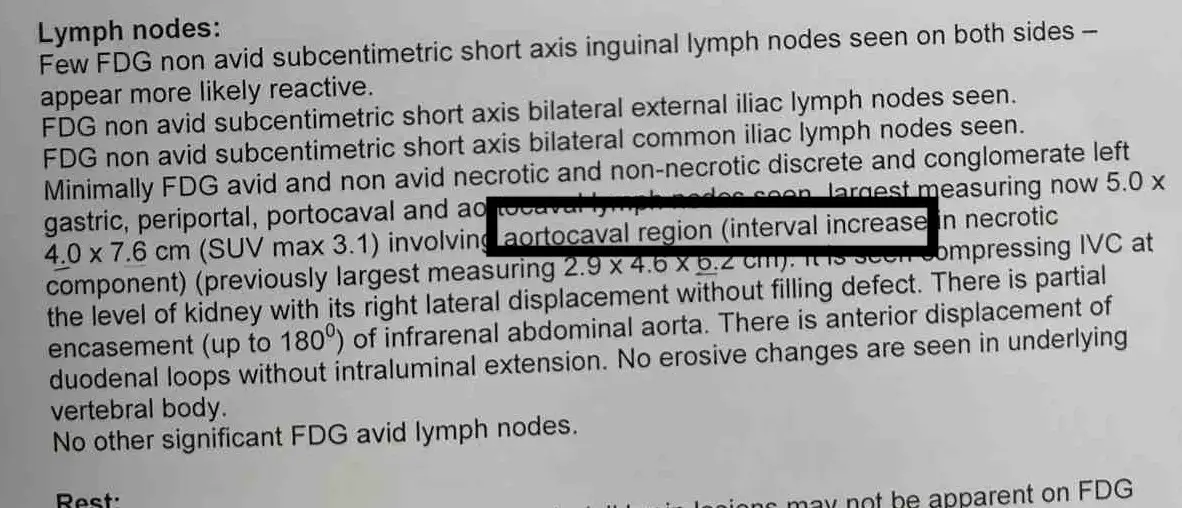
⦿ PET-CT showed enlarged aortocaval and portocaval lymph nodes with displacement of the inferior vena cava (IVC), partial encasement of the infrarenal abdominal aorta and close relation to the duodenum.
⦿ These lymph nodes lie immediately adjacent to the body’s major blood vessels. During surgery, meticulous dissection is required to remove all cancer while protecting the aorta, inferior vena cava, renal vessels and surrounding structures.
⦿ In selected patients with dense tumour adherence or vascular invasion, vascular resection and reconstruction may occasionally be required. For this reason, complex RPLND should ideally be performed in centres where vascular surgical expertise is immediately available. Careful pre-operative planning and multidisciplinary teamwork help achieve complete cancer clearance while minimizing complications.
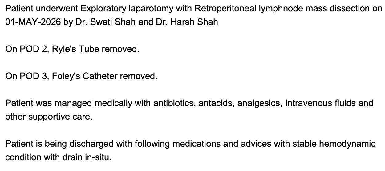
A recently managed Post-Chemotherapy Retroperitoneal Lymph Node Dissection (PC-RPLND) by Dr. Swati Shah, Uro Oncologist, Ahmedabad demonstrates how structured postoperative care supports recovery after major retroperitoneal cancer surgery.
The patient underwent Exploratory Laparotomy with Retroperitoneal Lymph Node Mass Dissection for a residual aortocaval, para-aortic and retropancreatic lymph node mass following chemotherapy for Right Testicular Malignant Germ Cell Tumour.
Recovery followed a planned postoperative pathway:
Modern recovery after RPLND is guided by Enhanced Recovery After Surgery (ERAS) principles, which focus on early mobilisation, optimal pain control, early nutrition and careful multidisciplinary postoperative care. These measures help reduce complications, improve bowel recovery and enable patients to return to normal daily activities sooner.
Take-home message:
Although Retroperitoneal Lymph Node Dissection is a major abdominal cancer operation, protocol-based perioperative care and ERAS principles have significantly improved recovery. With careful postoperative management, many patients can recover safely and resume normal activities much earlier than expected.
A recently managed testicular germ cell tumour case by Dr. Swati Shah, Uro Oncologist, Ahmedabad demonstrates an important principle in the treatment of Non-Seminomatous Germ Cell Tumour (NSGCT).
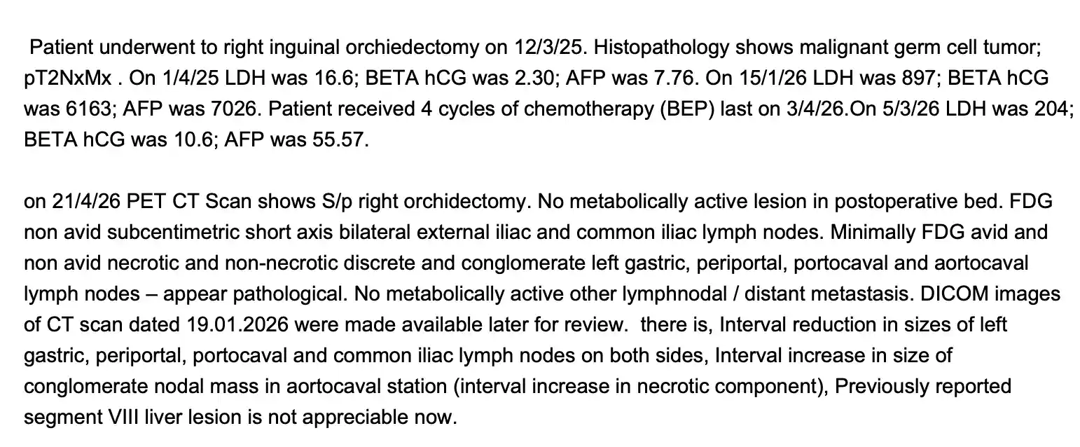
This patient was a known case of Right Testis Malignant Germ Cell Tumour (Yolk Sac + Embryonal) who underwent Right Inguinal Orchidectomy followed by 4 cycles of BEP chemotherapy. Before chemotherapy, Beta-hCG was 6163 IU/L, AFP was 7026 ng/mL and LDH was 897 U/L. After chemotherapy, all tumour markers showed an excellent decline. However, PET-CT still demonstrated a residual necrotic conglomerate aortocaval lymph node mass.
Many patients believe that normal tumour markers mean all cancer has disappeared.
Unfortunately, this is not always true.
Residual retroperitoneal masses after chemotherapy may contain:
⦿ Mature teratoma
⦿ Viable germ cell tumour
⦿ Necrosis or fibrosis
⦿ Tumour markers cannot differentiate between these possibilities.
Therefore, patients with residual retroperitoneal disease require further evaluation by a Uro Oncologist to determine whether Post-Chemotherapy Retroperitoneal Lymph Node Dissection (PC-RPLND) is indicated.
Take-home message:
Normal AFP, Beta-hCG and LDH are reassuring, but they do not exclude residual disease. Treatment decisions should always combine tumour markers, PET-CT findings and specialist surgical assessment.
A recently managed case by Dr. Swati Shah, Uro Oncologist, Ahmedabad highlights a rare but important phenomenon seen after chemotherapy for Non-Seminomatous Germ Cell Tumour (NSGCT).
Following 4 cycles of BEP chemotherapy, PET-CT demonstrated interval reduction in left gastric, periportal and portocaval lymph nodes, but the conglomerate aortocaval lymph node mass had increased in size with increased necrotic component.
The patient subsequently underwent Post-Chemotherapy Retroperitoneal Lymph Node Dissection (PC-RPLND).
Histopathology showed:
This illustrates an important concept.
Chemotherapy effectively destroys embryonal carcinoma and yolk sac tumour, but mature teratoma is resistant to chemotherapy and continues to require surgical removal.
Take-home message:
An enlarging or persistent retroperitoneal lymph node mass after chemotherapy does not always indicate treatment failure. It may represent mature teratoma, for which surgery—not additional chemotherapy—is the definitive treatment.
Histopathological examination revealed Deposits of Mature Cystic Teratoma composed of:
⦿ Mature cartilage
⦿ Sebaceous glands
⦿ Squamous epithelium
⦿ Gastric epithelium
⦿ Colonic epithelium
⦿ Salivary gland tissue
Importantly, there was no residual germ cell tumour component and no immature component.
Take-home message:
PET-CT identifies a residual mass, but only histopathology can determine whether it contains viable cancer, mature teratoma or fibrosis. This distinction directly influences whether further chemotherapy is necessary.
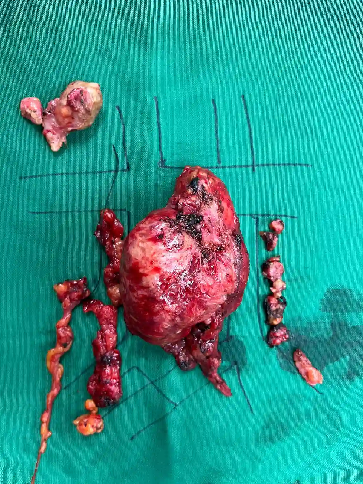
A recently managed Post-Chemotherapy Retroperitoneal Lymph Node Dissection (PC-RPLND) by Dr. Swati Shah, Uro Oncologist, Ahmedabad involved an approximately 8 × 8 cm aortocaval, precaval and para-aortic nodal mass, extending from the suprarenal region to the common iliac vessels, along with a 2 × 2 cm retropancreatic lymph node. The mass was densely adherent to the inferior vena cava (IVC) and abdominal aorta.
Surgery required:
Operations in this region are technically demanding because the tumour lies immediately adjacent to major blood vessels and vital structures.
In selected patients, extensive vascular involvement may require vascular resection and reconstruction. Therefore, these complex retroperitoneal cancer surgeries should ideally be planned in centres where vascular surgery support and multidisciplinary expertise are available if needed.
Take-home message:
Large aortocaval and retropancreatic lymph node masses after testicular cancer chemotherapy can often be removed safely with meticulous surgical planning, specialised retroperitoneal dissection and multidisciplinary support.
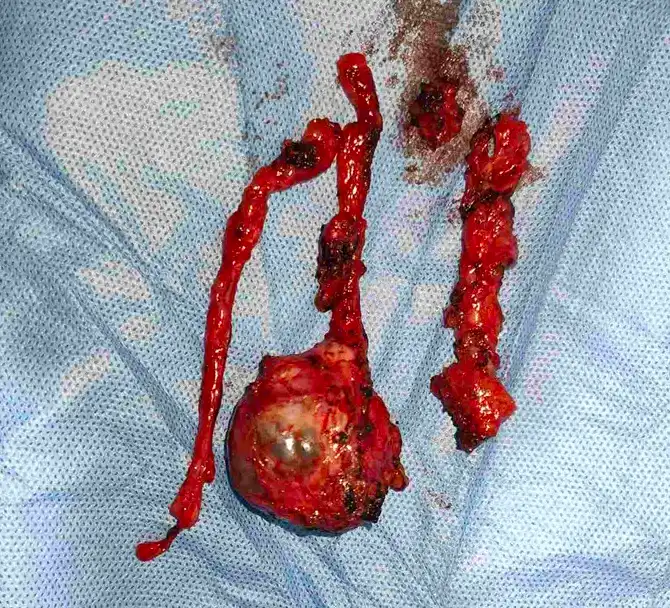
⦿ Recently managed case of recurrent non-seminomatous germ cell tumour (NSGCT) of the right testis in a 27-year-old man, previously treated with high inguinal orchiectomy and chemotherapy.
⦿ The patient had received 4 cycles of EP followed by 3 cycles of TIP chemotherapy. Follow-up CT showed a significant reduction in enhancement of the pre-caval lymph nodes with partial marginal reduction in size, but a residual retroperitoneal lymph-node mass persisted.
⦿ This is an important situation in testicular cancer: a residual retroperitoneal mass after chemotherapy needs careful assessment by a specialist experienced in testicular cancer, germ cell tumours and retroperitoneal lymph node dissection (RPLND).
⦿ In this case, the residual disease was not simply observed. The patient underwent exploratory laparotomy with complete retroperitoneal lymph node dissection, right external iliac lymph-node dissection and excision of residual right cord structures under the care of Dr. Swati Shah, Uro-Oncology & Surgical Oncology.
Keywords:
testicular cancer, non-seminomatous germ cell tumour, NSGCT, residual retroperitoneal lymph node, residual mass after chemotherapy, post-chemotherapy RPLND, testicular cancer surgery, Dr Swati Shah, uro-oncology.
Therefore, the clinical decision requires correlation of:
⦿ AFP, β-HCG and LDH
⦿ CT/PET-CT findings
⦿ previous histopathology
⦿ response to chemotherapy
⦿ characteristics and location of the residual retroperitoneal mass
The patient was subsequently taken for complete RPLND, allowing the residual mass to be removed and definitively examined by histopathology. Dr. Swati Shah managed the surgical component as a uro-oncology/surgical oncology case.
Keywords:
AFP in testicular cancer, residual mass after chemotherapy, NSGCT, non-seminomatous germ cell tumour, retroperitoneal lymph node, testicular cancer markers, post-chemotherapy RPLND, uro-oncology, Dr Swati Shah.
Recently managed complex retroperitoneal surgery for residual testicular cancer.
The operative findings in this 27-year-old patient illustrate why post-chemotherapy RPLND for testicular cancer can be technically demanding.
There was an approximately 3 × 3 cm solid-cystic mass in the para-aortic region, adherent to the aorta, beginning just below the left renal vessels. There were also enlarged inter-aortocaval lymph nodes and a right external iliac nodule.
The dissection required careful separation of the lymph-node tissue from:
⦿ aorta
⦿ left renal vessels
⦿ left gonadal vessels
⦿ ureter
⦿ inferior mesenteric artery (IMA)
A complete RPLND with full template dissection was performed, including paracaval, para-aortic, inter-aortocaval and retrocaval lymph-node tissue.
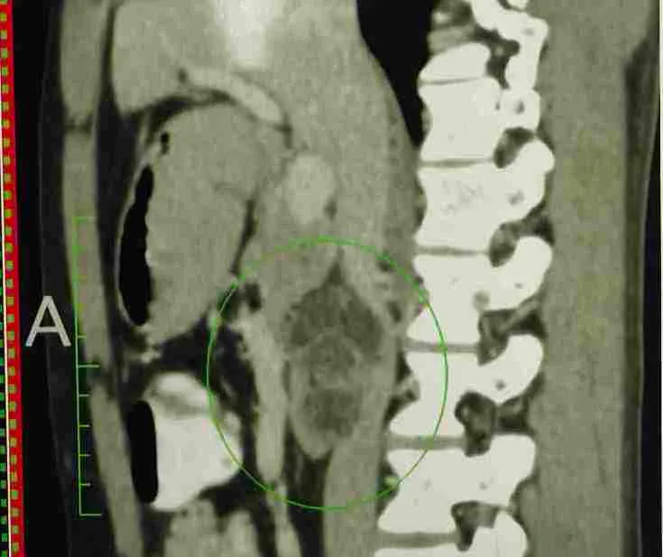
This type of surgery highlights the importance of precise retroperitoneal dissection and detailed knowledge of major vascular and urological anatomy in patients with residual testicular cancer.
In complex cases where the residual tumour is closely related to major vascular structures, appropriate vascular surgical support should also be available if vascular resection or reconstruction becomes necessary.
The surgery was performed by Dr. Swati Shah, Uro-Oncology & Surgical Oncology, with multidisciplinary operative support.
Keywords:
complex RPLND, retroperitoneal lymph node dissection, post-chemotherapy RPLND, para-aortic mass, inter-aortocaval lymph nodes, testicular cancer surgery, NSGCT, aortic dissection, renal vessels, uro-oncology, Dr Swati Shah.
The operation included:
⦿ Paracaval lymph-node dissection
⦿ Para-aortic lymph-node dissection
⦿ Inter-aortocaval lymph-node dissection
⦿ Retrocaval lymph-node dissection
⦿ Dissection of the residual right gonadal vessels and cord structures
⦿ Right external iliac lymph-node dissection
⦿ Removal of the right external iliac nodule
Keywords:
RPLND for testicular cancer, full-template RPLND, retroperitoneal lymph node dissection, post-chemotherapy NSGCT, residual retroperitoneal mass, para-aortic lymph nodes, inter-aortocaval lymph nodes, testicular cancer surgeon, uro-oncology, Dr Swati Shah.
Keywords:
residual retroperitoneal mass, NSGCT, testicular cancer, post-chemotherapy RPLND, pre-caval lymph nodes, IVC compression, para-aortic lymph nodes, complex RPLND, testicular cancer surgeon, uro-oncology, Dr Swati Shah.
The dissection therefore required meticulous identification and preservation of:
⦿ Aorta
⦿ IVC
⦿ Renal vessels
⦿ Gonadal vessels
⦿ Ureter
⦿ IMA
⦿ Duodenum and pancreas
The mass was carefully dissected from the aorta and left renal vessels, and a complete RPLND/full template dissection was performed.
Keywords:
testicular cancer RPLND, complex RPLND, vascular involvement, IVC compression, para-aortic mass, post-chemotherapy RPLND, retroperitoneal surgery, vascular surgeon, non-seminomatous germ cell tumour, uro-oncology, Dr Swati Shah.
Recently managed case of recurrent non-seminomatous germ cell tumour (NSGCT) of the right testis where definitive surgery was eventually required after progression of residual disease.
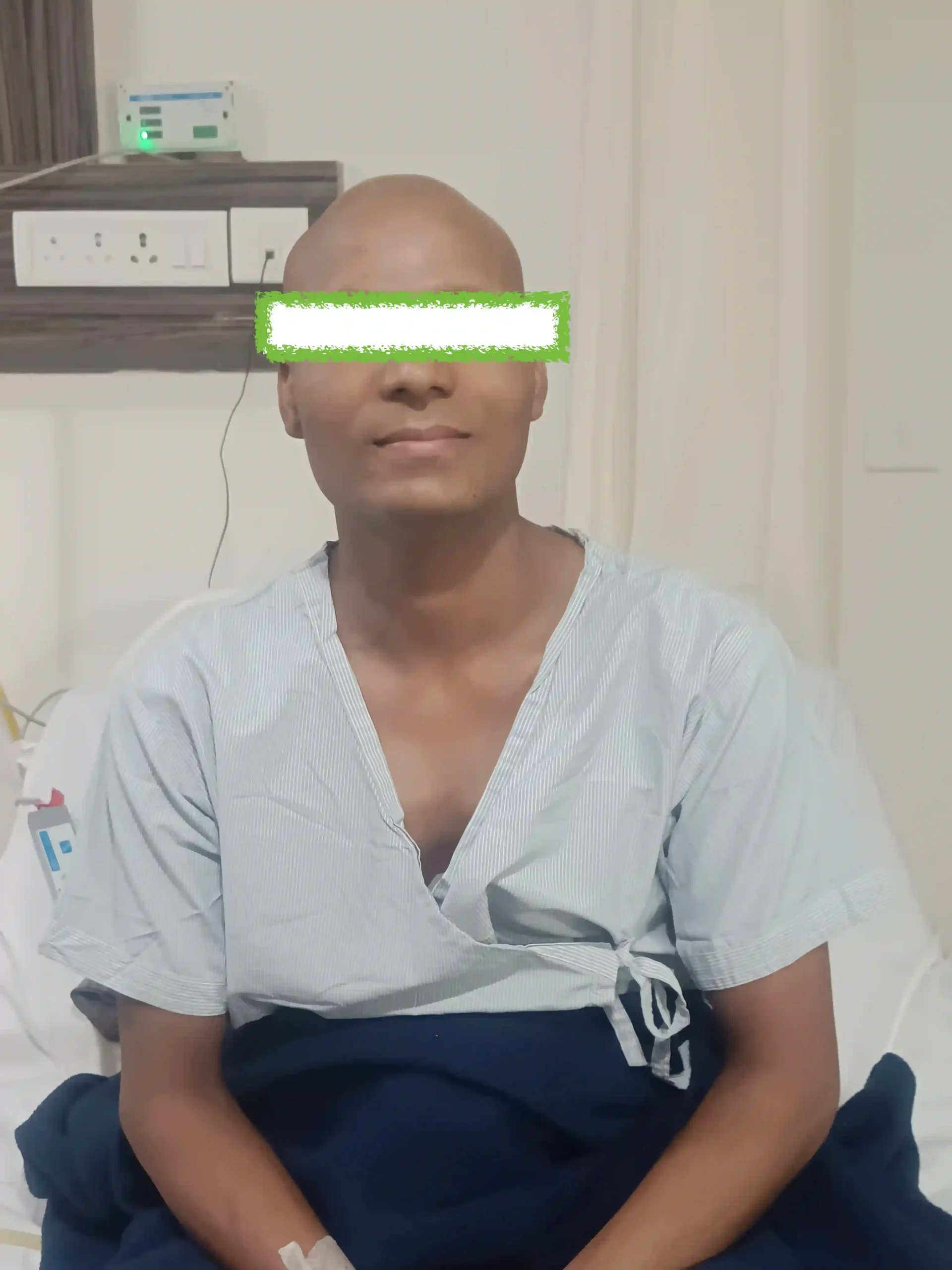
⦿ After the initial chemotherapy, the patient defaulted from planned treatment and did not want surgery. He was subsequently lost to follow-up and, over the following months, the retroperitoneal disease increased.
⦿ He later required further chemotherapy followed by definitive retroperitoneal surgery.
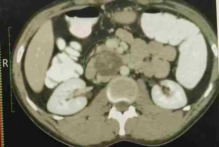
⦿ The subsequent CT demonstrated persistent/recurrent retroperitoneal disease, including pre-caval necrotic lymph nodes, a mass causing IVC compression/indentation, and disease along the right external iliac vessels.
⦿ This illustrates an important principle in testicular cancer: when a residual retroperitoneal mass requires surgical management, simply waiting for it to disappear may not be appropriate. The timing of chemotherapy, surgery and surveillance should be determined by a testicular cancer/uro-oncology specialist based on tumour markers, imaging and the type of germ cell tumour.
⦿ In this patient, definitive surgery ultimately involved complete retroperitoneal lymph node dissection, including para-aortic, paracaval, inter-aortocaval and retrocaval compartments.
⦿ Early specialist evaluation and adherence to the planned treatment pathway can prevent avoidable progression and make complex surgery more challenging than it needs to be.
Keywords:
recurrent testicular cancer, NSGCT, residual retroperitoneal mass, delayed surgery, post-chemotherapy RPLND, retroperitoneal lymph nodes, testicular cancer treatment, germ cell tumour, uro-oncology, Dr Swati Shah.
Recently managed case of recurrent right testicular non-seminomatous germ cell tumour with persistent retroperitoneal lymph-node disease after multiple chemotherapy regimens.
The final surgical procedure was not a limited removal of the largest node. The patient underwent complete RPLND with full template dissection.
The operative dissection included the:
Keywords:
complete RPLND, full-template RPLND, post-chemotherapy testicular cancer surgery, NSGCT, retroperitoneal lymph node dissection, para-aortic lymph nodes, inter-aortocaval nodes, paracaval nodes, retrocaval nodes, complex uro-oncology, Dr Swati Shah.
Swasth Parivar: every week, practical advice on recovery, diet and warning signs. No advertising. Stop any time.
Your number is used for these updates and for the clinic to reach you. It is never sold or passed on. To stop, reply STOP on WhatsApp.