
A recently managed case by Dr. Swati Shah, Uro Oncologist, Ahmedabad, involved a 66-year-old gentleman with Muscle Invasive Bladder Cancer (MIBC) who had a single left kidney following previous nephrectomy.
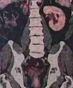
⦿ The TURBT biopsy confirmed High Grade Infiltrating Urothelial Carcinoma with lamina propria invasion and deep smooth muscle infiltration, confirming Muscle Invasive Bladder Cancer (MIBC). The preoperative PET-CT showed generalized thickening involving the left lateral wall of the urinary bladder, measuring about 45 mm, with no perivesical spread, no enlarged para-aortic or paracaval lymph nodes, no distant metabolically active disease, and importantly “Right kidney is not seen – H/O nephrectomy.”
⦿ Managing bladder cancer in patients with a single functioning kidney requires special expertise. Every step—from imaging, staging and treatment planning to surgery and urinary diversion—must be designed to preserve the remaining kidney while achieving complete cancer clearance.
Such patients should ideally be evaluated by an experienced Uro Oncologist who routinely manages Muscle Invasive Bladder Cancer, Radical Cystectomy, Ileal Conduit, and complex urinary reconstruction.
A recently managed Muscle Invasive Bladder Cancer (MIBC) case by Dr. Swati Shah, Uro Oncologist highlights an important principle in bladder cancer treatment.
The patient’s TURBT histopathology showed High Grade Infiltrating Urothelial Carcinoma , with deep smooth muscle infiltration , while PET-CT staged the disease as cT2N0M0 , with no distant metabolically active disease.
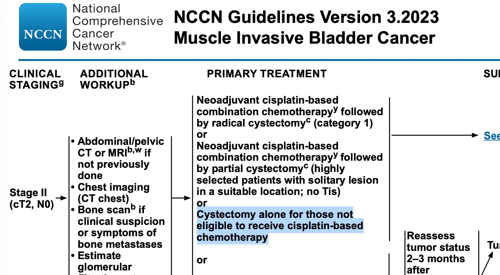
A recently managed Muscle Invasive Bladder Cancer (MIBC) case by Dr. Swati Shah, Uro Oncologist demonstrates why Radical Cystoprostatectomy remains the standard treatment for localized bladder cancer.
Preoperative evaluation showed:
⦿ High Grade Infiltrating Urothelial Carcinoma on TURBT
⦿ Deep smooth muscle infiltration confirming MIBC
⦿ PET-CT showing generalized thickening involving the left lateral wall of the urinary bladder without distant metastasis or perivesical spread.
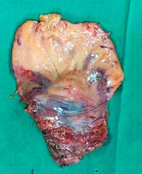
The patient underwent Robot Assisted Radical Cystoprostatectomy with Bilateral Pelvic Lymph Node Dissection and Ileal Conduit.
Final histopathology demonstrated High-grade Urothelial Carcinoma with Micropapillary Pattern (60–70%), measuring 4 × 3 cm, with negative surgical margins, while 21 lymph nodes were examined for accurate staging.
For Muscle Invasive Bladder Cancer, complete removal of the bladder along with appropriate pelvic lymph node dissection provides the best chance of long-term disease control in carefully selected patients.
A recently managed case by Dr. Swati Shah, Robotic Uro Oncologist, Ahmedabad highlights the role of Robot Assisted Radical Cystoprostatectomy in treating Muscle Invasive Bladder Cancer (MIBC).
The patient underwent Robot Assisted Radical Cystoprostatectomy with Bilateral Pelvic Lymph Node Dissection, Ileal Conduit and Umbilical Hernia Repair for High Grade Muscle Invasive Urothelial Carcinoma in a single kidney patient.
Robotic surgery offers several advantages in experienced hands:
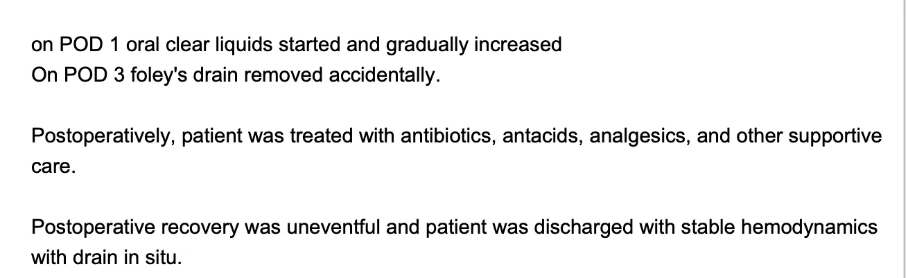
In this case, operative blood loss was approximately 300 ml, the patient remained hemodynamically stable, oral liquids were started on Postoperative Day 1, postoperative recovery was uneventful, and the patient was discharged in a stable condition.
For appropriately selected patients, Robotic Radical Cystectomy has become an important option for managing Muscle Invasive Bladder Cancer, particularly in high-volume centres with expertise in advanced robotic uro-oncology surgery.
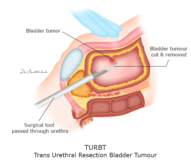
A recently managed case by Dr. Swati Shah, Uro Oncologist, involved an 82-year-old gentleman who presented with painless haematuria (blood in urine). CT IVP showed a heterogeneously mildly enhancing luminal protruding mass arising from the left lateral urinary bladder wall, while PET-CT demonstrated an FDG avid enhancing polypoidal soft tissue lesion with no evidence of distant metastasis.
Many patients believe that advanced age alone makes bladder cancer surgery unsafe. In reality, treatment decisions depend more on overall fitness, associated medical conditions, tumour stage and anaesthetic assessment than on chronological age.
Following multidisciplinary evaluation, the patient underwent cystoscopy with Transurethral Resection of Bladder Tumour (TURBT). He recovered well after surgery and was discharged in stable condition.
If you or your family member has blood in urine, bladder mass, urinary bladder tumour, or suspected bladder cancer, early evaluation by an experienced uro oncologist is important. Even patients in their 80s can often receive effective treatment.
In another recently managed bladder cancer case by Dr. Swati Shah, Uro Oncologist, the patient’s CT IVP demonstrated several classical findings suggestive of bladder malignancy.
The report described a heterogeneously mildly enhancing luminal protruding mass lesion arising from the lower portion of the left lateral urinary bladder wall, along with adjacent focal enhancing mural thickening. The radiologist concluded that these findings raised the possibility of primary neoplastic etiology and recommended further evaluation with cystoscopy and histopathology.
CT also helps evaluate:
However, CT scan alone cannot confirm bladder cancer. Tissue diagnosis through TURBT and histopathology is always required before planning definitive treatment.
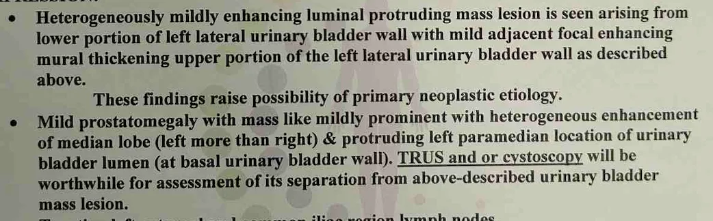
Patients searching for CT scan showing bladder mass, bladder wall thickening, bladder tumour CT findings or bladder cancer diagnosis should undergo evaluation by a dedicated uro oncologist.
A recently managed bladder cancer case by Dr. Swati Shah, Uro Oncologist, illustrates the value of PET-CT in treatment planning.
The PET-CT reported an FDG avid enhancing polypoidal soft tissue lesion based on the left lateral wall of the urinary bladder, along with a suspicious nodular lesion on the anterior bladder wall, while importantly showing no FDG avid pelvic, retroperitoneal or mesenteric lymph nodes and no distant metastatic disease.
PET-CT helps answer several important questions:
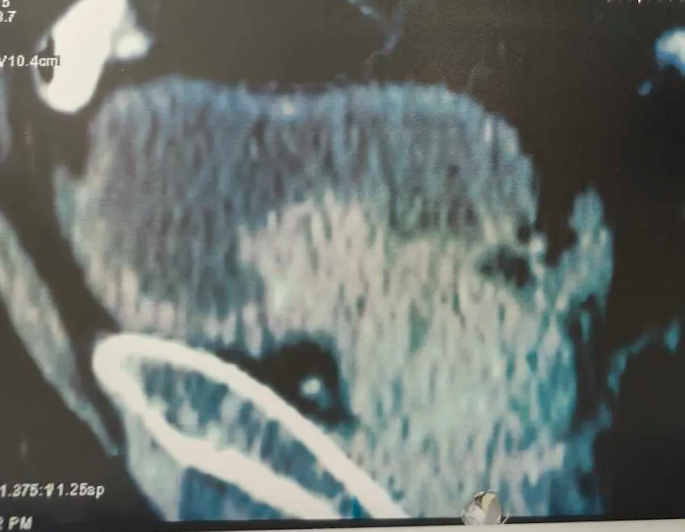
PET-CT findings must always be correlated with cystoscopy, TURBT and histopathology before deciding the final treatment plan.
Patients searching for PET scan for bladder cancer, bladder cancer staging, FDG avid bladder lesion or urothelial carcinoma PET findings should consult an experienced uro oncologist for comprehensive evaluation.
A recently managed case by Dr. Swati Shah, Uro Oncologist, involved an 82-year-old gentleman with a large 4 × 4 cm left lateral wall bladder mass.
During cystoscopy, the tumour was carefully assessed. Both ureteric openings were clearly identified and preserved before performing a complete Transurethral Resection of Bladder Tumour (TURBT). Separate superficial and deep bladder tumour specimens were sent for histopathological examination.
Histopathology confirmed High-grade urothelial carcinoma with focal micropapillary pattern, with lamina propria invasion as well as muscularis propria (detrusor muscle) invasion, establishing the diagnosis of muscle-invasive bladder cancer (MIBC).
A properly performed TURBT is not just a biopsy—it provides:
For patients with bladder tumour, bladder mass, urothelial carcinoma or muscle-invasive bladder cancer, a meticulous TURBT performed by an experienced uro oncologist is the foundation of successful treatment.
A recently managed bladder cancer case by Dr. Swati Shah, Uro Oncologist, reminds us that bladder cancer is not only a disease of men.
This 42-year-old lady presented with blood in urine (hematuria) for nearly 20 days. Ultrasound showed a urinary bladder mass, and she underwent further evaluation followed by cystoscopy and transurethral resection of bladder tumour (TURBT).
Many women mistake blood in urine as:
In India, bleeding may also occur because of cervical cancer involving the urinary bladder, but primary bladder cancer should always be ruled out, especially when bleeding is painless.
Persistent or recurrent hematuria in females should never be ignored. Early diagnosis allows timely treatment and better outcomes.
A recently managed case by Dr. Swati Shah, Uro Oncologist, highlights the importance of a simple ultrasound.
The ultrasound report demonstrated:
When evaluating a bladder tumour on ultrasound, experienced uro-oncologists look for:
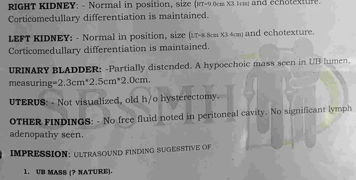
Although ultrasound raises suspicion, cystoscopy with biopsy or TURBT is essential for definitive diagnosis.
A recently managed bladder cancer case by Dr. Swati Shah, Uro Oncologist, demonstrates why a proper Transurethral Resection of Bladder Tumour (TURBT) is critical.
During cystoscopy:
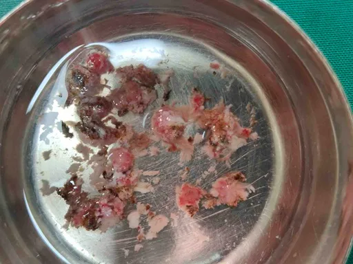
Final histopathology confirmed:
A deep muscle biopsy is extremely important because the presence or absence of detrusor muscle invasion determines whether the cancer is non-muscle-invasive bladder cancer (NMIBC) or muscle-invasive bladder cancer (MIBC) and directly influences further treatment.
A recently managed young female bladder cancer case by Dr. Swati Shah, Uro Oncologist, raises an important question many patients ask.
After TURBT, the final pathology showed:
In carefully selected young patients with disease confined to the bladder and suitable kidney function, treatment may include:
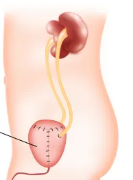
Not every patient is a candidate for a neobladder. Age, tumour location (especially near the bladder neck or urethra), kidney function, continence status and overall health are carefully evaluated before deciding the most appropriate urinary diversion.
Proper evaluation by an experienced uro-oncologist helps determine whether neobladder reconstruction is a safe and feasible option.
A recently managed case by Dr. Swati Shah, Uro Oncologist, highlights why Pelvic Lymph Node Dissection (PLND) is an essential part of surgery for Muscle Invasive Bladder Cancer (MIBC).
The preoperative PET-CT showed no definite enlarged para-aortic, paracaval or portal lymph nodes, and no distant metabolically active disease. However, surgery still included a Bilateral Pelvic Lymph Node Dissection because microscopic lymph node spread cannot always be detected on imaging.
Final histopathology demonstrated:
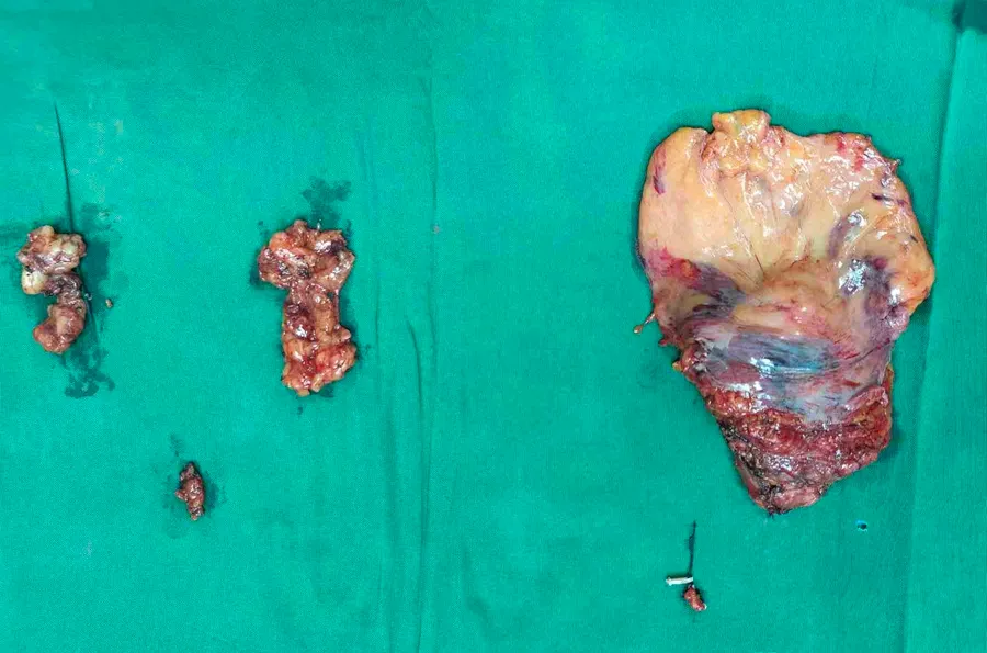
This case shows why Pelvic Lymph Node Dissection provides accurate staging, guides further treatment decisions, and improves oncological management. Even when PET-CT appears reassuring, lymph node removal remains an important component of surgery for Muscle Invasive Bladder Cancer.
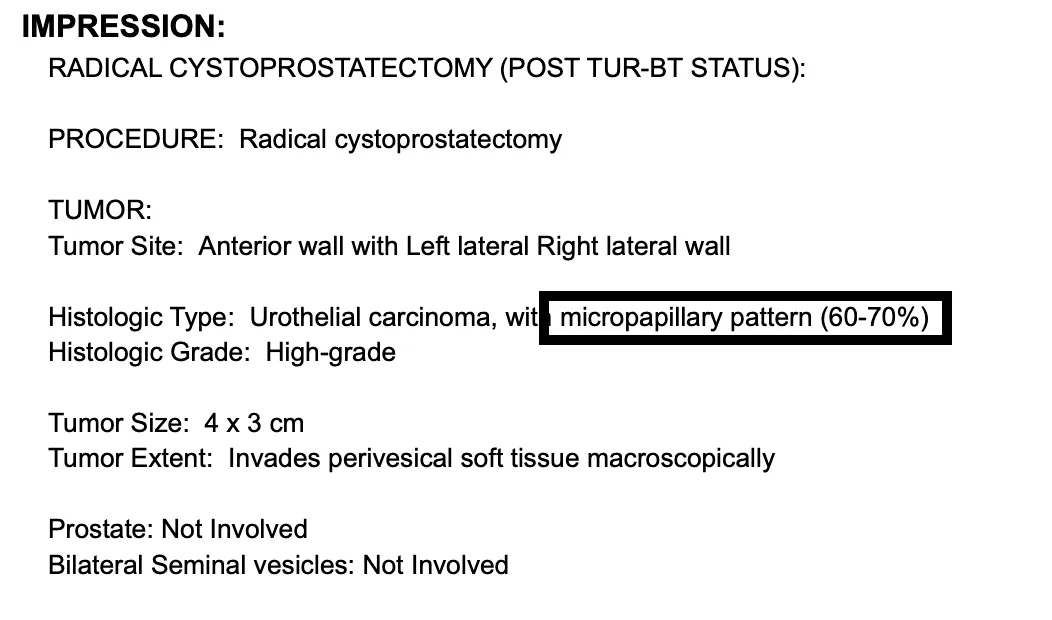
A recently managed case by Dr. Swati Shah, Uro Oncologist, demonstrated an uncommon but important subtype of bladder cancer.
Initial TURBT reported High Grade Infiltrating Urothelial Carcinoma with deep smooth muscle infiltration, confirming Muscle Invasive Bladder Cancer (MIBC).
However, the final specimen after Robot Assisted Radical Cystoprostatectomy revealed:
Certain aggressive variants, such as Micropapillary Urothelial Carcinoma, may not be fully recognised on the initial TURBT specimen because only a limited portion of the tumour is sampled.
This highlights why definitive surgery and complete histopathological examination remain essential in many patients with Muscle Invasive Bladder Cancer.
A recently managed Muscle Invasive Bladder Cancer case by Dr. Swati Shah, Uro Oncologist, reinforces an important concept.
The PET-CT reported:
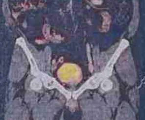
Despite these reassuring findings, final pathology after Radical Cystoprostatectomy showed:
PET-CT is an excellent staging investigation but cannot detect every microscopic focus of disease.
For patients with High Grade Muscle Invasive Urothelial Carcinoma, definitive surgery remains the cornerstone of treatment even when imaging does not show advanced disease.
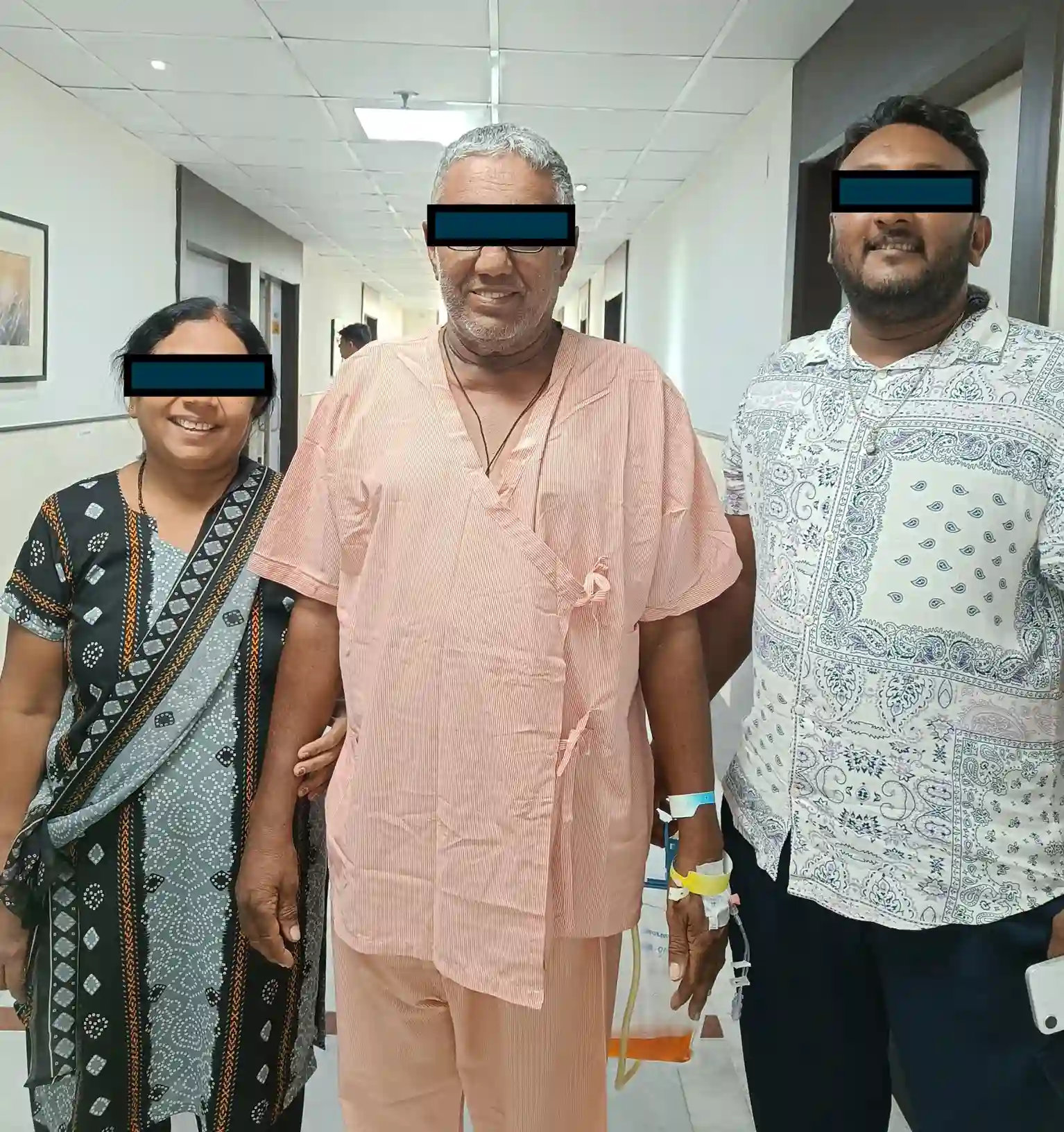
A recently managed case by Dr. Swati Shah, Robotic Uro Oncologist, demonstrates that recovery after major bladder cancer surgery can be smoother with protocol-based perioperative care.
The patient underwent Robot Assisted Radical Cystoprostatectomy with Bilateral Pelvic Lymph Node Dissection, Ileal Conduit and Umbilical Hernia Repair for Muscle Invasive Bladder Cancer (MIBC).
Recovery milestones included:
Today, even after Radical Cystectomy, Pelvic Lymph Node Dissection, and Ileal Conduit, many patients can recover well with Enhanced Recovery After Surgery (ERAS) principles, experienced anaesthesia, specialised nursing, physiotherapy, nutritional support, and coordinated multidisciplinary care provided by a dedicated Uro Oncology team.
A recently managed case by Dr. Swati Shah, Uro Oncologist, highlights why histopathology (HPE) after Transurethral Resection of Bladder Tumour (TURBT) is the most important investigation in bladder cancer.
Although CT IVP and PET-CT strongly suggested a bladder tumour, the final diagnosis depended on microscopic examination of the resected tissue.
Histopathology showed:
These findings confirmed muscle-invasive bladder cancer (MIBC) and determined the need for definitive treatment such as radical cystectomy with urinary diversion, rather than repeated TURBT alone.
Patients searching for high-grade urothelial carcinoma, muscle-invasive bladder cancer, bladder biopsy report, lamina propria invasion or detrusor muscle invasion should understand that the histopathology report—not imaging alone—determines the next step in treatment.
A recently managed case by Dr. Swati Shah, Uro Oncologist, demonstrates that age alone should not prevent treatment of bladder cancer.
This 82-year-old gentleman underwent complete TURBT for a bladder tumour. With careful anaesthesia, pain management, early mobilisation and protocol-based postoperative care, recovery was smooth.
Most patients undergoing TURBT can expect:
In this case, the patient was mobilised on Day 1 and discharged on Day 2 after an uneventful recovery.
Patients often search for recovery after bladder tumour surgery, TURBT recovery time, hospital stay after TURBT, bladder cancer surgery in elderly patients and should know that recovery is generally much quicker than expected.
A recently managed bladder cancer case by Dr. Swati Shah, Uro Oncologist, began with a symptom that many patients ignore—painless haematuria (blood in urine).
Investigations subsequently revealed:
Blood in urine may occur only once and disappear, but it should never be ignored, especially in older adults or smokers.
Common warning symptoms include:
Early consultation with a uro oncologist allows diagnosis while the disease is still potentially curable.
A recently managed female bladder cancer case by Dr. Swati Shah, Uro Oncologist, highlights an important fact—not every bladder cancer patient has a history of smoking or tobacco use.
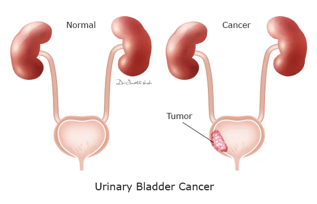
This patient had:
Smoking remains the biggest risk factor for bladder cancer, but other causes include:
To reduce the risk of bladder cancer:
Early diagnosis often allows more effective treatment.
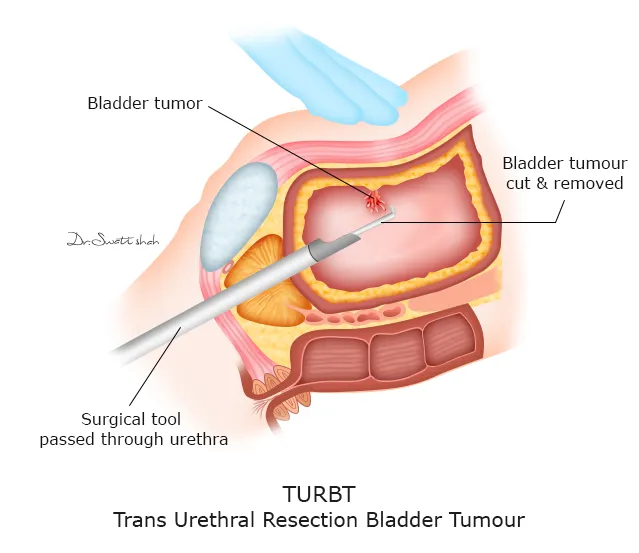
A recently managed bladder cancer case by Dr. Swati Shah, Uro Oncologist, demonstrates why the histopathology report after TURBT is more important than imaging alone.
The TURBT specimen was submitted separately as:
Final histopathology reported:
These findings confirmed muscle-invasive bladder cancer (MIBC).
Whether a patient requires:
depends primarily on the histopathology findings, not just ultrasound, CT or PET-CT.
Obtaining an adequate deep muscle specimen during TURBT is therefore essential.
A recently managed bladder tumour case by Dr. Swati Shah, Uro Oncologist, shows that recovery after Transurethral Resection of Bladder Tumour (TURBT) is generally rapid.
After surgery:
Most patients undergoing uncomplicated TURBT can:
Recovery is usually much easier than patients anticipate.
A recently managed case by Dr. Swati Shah, Uro Oncologist, reminds us that muscle-invasive bladder cancer is treatable when diagnosed appropriately and managed in time.
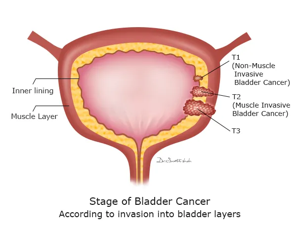
This patient initially presented with:
Following complete evaluation:
For patients with muscle-invasive bladder cancer, further management may include:
Early referral to an experienced uro-oncologist allows treatment planning before the disease progresses, improving the chances of long-term cancer control and quality of life.
⦿ Recently managed at Apollo Hospital, Ahmedabad, a 55-year-old man presented with lower urinary tract symptoms (LUTS) and straining. He had undergone TURBT (transurethral resection of bladder tumour), and histopathology showed urachal adenocarcinoma.
⦿ Not every bladder mass is a typical urothelial carcinoma.
⦿ The commonest bladder cancer is urothelial carcinoma, but a bladder mass can occasionally represent a different tumour type such as adenocarcinoma arising from the urachus.
⦿ In this case, the pathology established adenocarcinoma of the urinary bladder, and the clinical and operative findings supported a urachal origin. Therefore, simply treating the lesion as a routine superficial bladder tumour would not have been adequate.
⦿ The distinction between urothelial carcinoma vs bladder adenocarcinoma vs urachal adenocarcinoma is important because the surgical approach can be very different.
⦿ The patient subsequently underwent definitive surgery with partial cystectomy and bilateral pelvic lymph-node dissection, along with excision of the urachal remnant.
Key message:
When a bladder mass is diagnosed after TURBT, the exact histological type and anatomical origin should be established before deciding the definitive treatment.
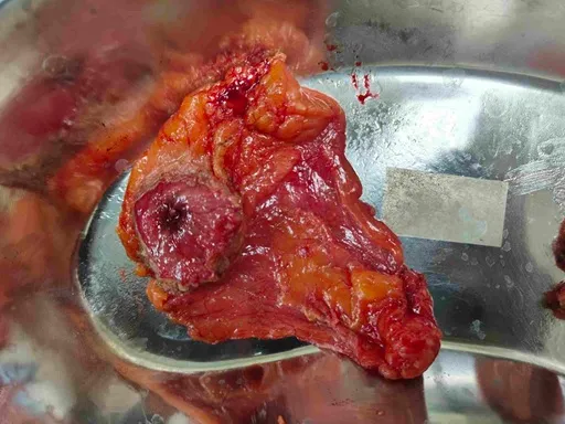
Recently managed at Apollo Hospital, Ahmedabad, a 55-year-old man with urachal adenocarcinoma of the urinary bladder underwent partial cystectomy rather than removal of the entire bladder.
⦿ The operation included removal of the tumour with approximately a 2-cm normal bladder margin.
⦿ The surgical note describes mobilisation of the urachal remnant from the umbilicus towards the bladder, excision of the bladder mass with normal surrounding tissue, and double-layer closure of the bladder.
⦿ Why is this important?
⦿ For selected bladder cancers, particularly appropriately located urachal tumours, complete tumour removal does not necessarily mean removing the entire bladder.
The aim is:
In this patient, final histopathology showed all surgical margins negative for invasive tumour.
Key message:
Partial cystectomy is not simply a “smaller operation.” It is a carefully selected bladder-preserving cancer surgery where achieving complete tumour removal with clear margins remains the priority.
⦿ A bladder mass located near the bladder dome needs careful evaluation because it may arise from the urachus.
⦿ Recently managed at Apollo Hospital, Ahmedabad, a patient with urachal adenocarcinoma underwent partial cystectomy with removal of the urachal remnant.
⦿ During surgery, the urachal remnant was mobilised from the umbilicus towards the bladder, followed by excision of the bladder tumour with a normal tissue margin.
⦿ The reason is anatomical.
⦿ The urachus is a remnant of the embryological connection between the bladder and umbilicus. Tumours arising from this structure are known as urachal cancers, and the surgical plan therefore needs to address not only the visible bladder mass but also the involved urachal tract.
⦿ In this case, the bladder lesion was excised with approximately a 2-cm normal bladder margin, followed by bladder closure.
⦿ Final histopathology confirmed adenocarcinoma, moderately differentiated (G2).
Key message:
A bladder dome mass is not always a routine urothelial bladder cancer. Recognising a possible urachal origin can change the extent and type of bladder cancer surgery.
⦿ Recently managed at Apollo Hospital, Ahmedabad, a 55-year-old man with urachal adenocarcinoma underwent partial cystectomy with bilateral pelvic lymph-node dissection.
⦿ The lymph nodes were removed on both sides to provide accurate pathological staging and determine whether the bladder cancer had spread to regional lymph nodes.
⦿ The final histopathology was reassuring in this respect:
⦿ 15 pelvic lymph nodes were examined — 0/15 showed metastatic tumour.
⦿ The tumour itself was a 0.7 × 0.5 cm adenocarcinoma, moderately differentiated (G2), but importantly it showed microscopic invasion into perivesical soft tissue.
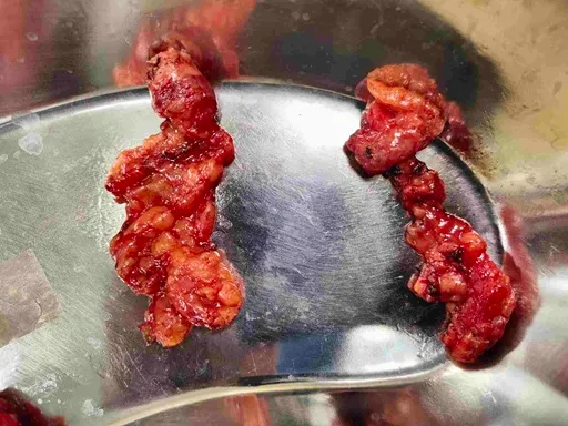
⦿ The final pathological stage was:
⦿ pT3a pN0
⦿ This demonstrates an important principle in bladder cancer: tumour size alone does not determine the seriousness of the disease.
⦿ The depth of invasion, surgical margins and lymph-node status all contribute to accurate staging and further treatment planning.
Key message:
During bladder cancer surgery, pelvic lymph-node dissection is not merely an additional procedure. It provides critical information about cancer spread and helps determine the final stage of disease.
⦿ Recently managed case of high-grade urothelial carcinoma involving the urethra in a patient previously treated for muscle-invasive bladder cancer.
⦿ The patient had undergone robotic radical cystectomy + partial prostatectomy + bilateral pelvic lymph-node dissection + neobladder reconstruction in February 2025.
⦿ He subsequently developed blood in urine. Cystoscopy showed a urethral growth, and biopsy of the urethral lesion on 08/04/2026 showed high-grade invasive urothelial carcinoma.
⦿ The CT abdomen and pelvis showed mild bilateral hydroureter, with no pelvic lymphadenopathy. A 17 × 18 mm liver lesion was considered possibly a hemangioma, rather than definite metastatic disease.
⦿ Because of the urethral disease, total urethrectomy with permanent suprapubic catheterisation was performed.
⦿ The OT findings were important: the urethra was thickened and there were two lesions in the anterior urethra, approximately 4 cm and 10 cm from the meatus. The neobladder and anastomotic site were normal.
⦿ Final histopathology of the completely submitted 13-cm urethrectomy specimen showed no residual tumour. The final pathology report therefore records the stage according to the previous biopsy: high-grade urothelial carcinoma involving subepithelial connective tissue, pT1a.
⦿ This case highlights why hematuria after bladder cancer treatment should not be ignored, even when the bladder has already been removed.
Key message:
Urethral cancer | Urethral recurrence | High-grade urothelial carcinoma | Urethral growth | Hematuria after cystectomy | Total urethrectomy | Neobladder | Bladder cancer recurrence | Uro-oncology
⦿ Recently managed case of urethral cancer diagnosed after a patient presented with blood in urine.
⦿ This case demonstrates an important principle: a visible urethral growth requires tissue diagnosis before definitive treatment is planned.
⦿ The patient had a history of muscle-invasive bladder cancer, previously treated with radical cystectomy and neobladder reconstruction. When he developed blood in urine, cystoscopy was performed and a urethral lesion was identified.
⦿ Biopsy showed:
⦿ High-grade invasive urothelial carcinoma.
⦿ The pathology demonstrated involvement of the subepithelial connective tissue, with the previous biopsy staged as pT1a.
⦿ The biopsy was therefore critical—not simply to confirm that the lesion was malignant, but to establish the histological type, grade and depth of invasion, which helped guide definitive surgery.
⦿ In the subsequent urethrectomy specimen, the entire 13-cm tubular specimen was submitted for examination and no residual tumour was identified.
⦿ For patients with a urethral mass, urethral bleeding or unexplained hematuria, evaluation may include clinical examination, cystoscopy/urethroscopy, biopsy and appropriate imaging, depending on the clinical situation.
Key message:
Urethral cancer | Urethral tumour | Urethral mass | Urethral growth | Urothelial carcinoma | High-grade urothelial carcinoma | Urethral biopsy | Cystoscopy | Hematuria | Uro-oncologist
⦿ Recently managed case demonstrating the importance of histopathology in urethral cancer.
⦿ The patient had previously undergone radical cystectomy with neobladder reconstruction for muscle-invasive bladder cancer and later developed a urethral growth associated with blood in urine.
⦿ Cystoscopy-guided biopsy showed:
⦿ High-grade invasive urothelial carcinoma with subepithelial connective tissue involvement — pT1a.
⦿ The subsequent surgical specimen consisted of a 13-cm urethrectomy specimen, which was entirely submitted for histopathological examination.
⦿ Interestingly, the final urethrectomy specimen showed no residual tumour. The pathology report specifically states that staging was therefore based on the previous biopsy.
⦿ This is an important lesson for patients: the biopsy and the final surgical specimen answer different questions.
⦿ Therefore, treatment decisions in urethral cancer should be based on clinical findings + imaging + biopsy histology + definitive surgical pathology, rather than on imaging alone.
Key message:
Urethral urothelial carcinoma | High-grade urothelial carcinoma | Urethral biopsy | Histopathology | pT1a urethral cancer | Urethral tumour treatment | Uro-oncology
⦿ Recently managed urethral cancer requiring total urethrectomy in a patient with a previous neobladder.
⦿ The preoperative diagnosis was carcinoma urethra in a patient with previous muscle-invasive bladder cancer treated with radical cystoprostatectomy and neobladder reconstruction.
⦿ The operation was planned after biopsy confirmed high-grade invasive urothelial carcinoma.
⦿ At surgery, the urethra was thickened, with two anterior urethral lesions—approximately 4 cm and 10 cm from the meatus.
⦿ A total urethrectomy with permanent SPC was performed through a vertical midline perineal approach.
⦿ The operative steps included:
⦿ The final urethrectomy specimen showed no residual tumour, while the previous biopsy established high-grade urothelial carcinoma, pT1a.
⦿ The postoperative recovery was uneventful. The patient was ambulatory and was discharged on postoperative day 2 with the drain in situ.
Key message:
Total urethrectomy | Urethral cancer surgery | Urethral urothelial carcinoma | Neobladder | Radical cystectomy | Permanent SPC | Perineal urethrectomy | Uro-oncological surgery | Bladder cancer surgery
⦿ Recently managed at Apollo Hospital, Ahmedabad, this patient had initially presented with a bladder mass and underwent TURBT.
⦿ The important question after TURBT was not only “Is this bladder cancer?” but also:
⦿ “What type of bladder cancer is this?”
⦿ Histopathology demonstrated adenocarcinoma, rather than the more common urothelial carcinoma, with features supporting a urachal origin.
⦿ This distinction is important because the management of urothelial bladder cancer and urachal adenocarcinoma is not identical.
⦿ In this case, the diagnosis led to definitive cancer surgery with:
⦿ Final pathology showed moderately differentiated adenocarcinoma (G2) with perivesical soft-tissue invasion, staged pT3a pN0.
Key message:
A bladder tumour should not be treated based on imaging alone. TURBT and histopathology establish the tumour type and depth of invasion, which are essential for planning definitive bladder cancer treatment.
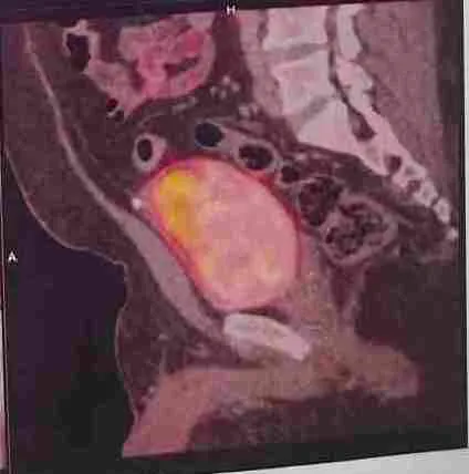
Recently managed at Apollo Hospital, Ahmedabad, a patient with urachal adenocarcinoma presenting as a bladder mass underwent bladder-preserving cancer surgery.
The tumour was removed by partial cystectomy, with approximately a 2-cm normal bladder margin.
The final histopathology demonstrated:
⦿ Moderately differentiated adenocarcinoma (G2)
⦿ Microscopic invasion into perivesical soft tissue
⦿ All margins negative for invasive tumour
⦿ Bladder base negative
⦿ Circumferential margins negative
⦿ 0/15 pelvic lymph nodes involved
⦿ The operation therefore achieved an R0 resection—complete removal of the visible tumour with no tumour identified at the examined surgical margins.
In cancer surgery, removing the visible mass is only part of the objective. The surgeon must also achieve an adequate margin of normal tissue while preserving as much useful bladder function as oncologically safe.
Key message:
For bladder cancer and urachal cancer surgery, clear surgical margins are one of the most important indicators of the quality and completeness of tumour removal.
Recently managed at Apollo Hospital, Ahmedabad, a patient with a bladder mass underwent TURBT (transurethral resection of bladder tumour).
TURBT is an important first procedure for many patients with suspected bladder cancer because it allows the surgeon to:
In this case, TURBT provided the crucial diagnosis of urachal adenocarcinoma.
That changed the treatment pathway.
Instead of treating the patient as a routine superficial urothelial bladder cancer, definitive surgery was planned with partial cystectomy, excision of the urachal remnant and bilateral pelvic lymph-node dissection.
Final pathology demonstrated perivesical soft-tissue invasion (pT3a).
Key message:
TURBT can be the crucial diagnostic and initial treatment procedure for a bladder mass, but some tumours require definitive bladder cancer surgery after the histopathology is available.
⦿ Recently managed at Apollo Hospital, Ahmedabad, this case highlights why size alone should never be used to judge the seriousness of a bladder tumour.
⦿ The final specimen contained a tumour measuring only approximately 0.7 × 0.5 cm.
⦿ Yet histopathology showed moderately differentiated adenocarcinoma (G2) with microscopic invasion into the perivesical soft tissue.
⦿ The final pathological stage was: pT3a pN0
⦿ The patient had undergone partial cystectomy with excision of the urachal remnant and bilateral pelvic lymph-node dissection. All examined margins were negative and 0/15 lymph nodes contained metastatic tumour.
⦿ So, how can a tumour that appears small still be significant?
⦿ Because bladder cancer assessment depends on much more than dimensions. Important factors include:
⦿ Tumour type → depth of invasion → perivesical involvement → lymph-node status → surgical margins.
⦿ This is particularly relevant when evaluating an unusual bladder mass, where distinguishing urothelial carcinoma from adenocarcinoma/urachal adenocarcinoma can completely change the treatment approach.
Key message:
Even a small bladder tumour can require major oncological treatment if pathology demonstrates invasive disease.
⦿ Recently managed case of a patient who had previously undergone radical cystoprostatectomy with neobladder reconstruction for muscle-invasive bladder cancer (MIBC).
⦿ A neobladder is a form of urinary reconstruction in which a segment of bowel is used to create a new reservoir for urine after the urinary bladder has been removed.
⦿ In this patient, the subsequent urethral cancer surgery demonstrated that the neobladder and anastomotic site were normal. Therefore, although the urethra required complete removal, the neobladder itself could be preserved.
⦿ This highlights an important aspect of urinary reconstruction: management after radical cystectomy is not always simply about the bladder being removed. The type of urinary diversion, urethral status and relationship of any subsequent tumour to the neobladder all influence further treatment.
⦿ Patients considering bladder cancer surgery often ask about neobladder vs ileal conduit, urinary reconstruction after cystectomy, continence and life after bladder removal.
⦿ The appropriate urinary diversion depends on the patient’s cancer, anatomy, kidney function, general health and suitability for reconstruction.
Key message:
Bladder cancer | Muscle-invasive bladder cancer | Radical cystectomy | Neobladder | Urinary reconstruction | Bladder removal | Uro-oncology | Dr Swati Shah
⦿ Recently managed case of urethral cancer in a patient who had previously undergone radical cystoprostatectomy with neobladder reconstruction for muscle-invasive bladder cancer.
⦿ One of the important surgical questions was whether the urethral tumour had involved the neobladder or the anastomotic site.
⦿ At operation, there was a thickened urethra with two anterior urethral lesions, approximately 4 cm and 10 cm from the meatus.
⦿ Importantly: The anastomotic site and neobladder were normal.
⦿ This allowed the surgical team to perform total urethrectomy while preserving the neobladder.
⦿ Under cystoscopic guidance, the proximal urethra was divided at the junction with the neobladder, and the neobladder was subsequently closed in layers.
⦿ This case demonstrates why the relationship between the urethral tumour and urinary reconstruction must be assessed carefully before surgery.
⦿ When urethral cancer occurs in a patient with a neobladder, the operation needs to address the cancer while preserving unaffected urinary reconstruction whenever oncologically and technically possible.
Key message:
Urethral cancer | Urethral tumour | Neobladder | Radical cystectomy | Total urethrectomy | Urinary reconstruction | Bladder cancer | Uro-oncology | Dr Swati Shah
⦿ Recently managed case of urethral cancer requiring total urethrectomy in a patient with previous radical cystoprostatectomy and neobladder reconstruction.
⦿ The operation was performed through a vertical midline perineal incision in the lithotomy position.
⦿ The urethra was circumferentially mobilised after division of the Colles fascia and bulbospongiosus muscle. The central tendon of the perineum was divided to separate the bulb from the rectum.
⦿ Distally, the urethra was carefully dissected away from the corpora cavernosa and circumferentially dissected from the meatus.
⦿ Importantly, the penis was preserved.
⦿ Proximally, the uropelvic diaphragm was divided, and under cystoscopic guidance the urethra was divided at the junction with the neobladder. The neobladder was then closed in layers.
⦿ This illustrates the complexity of urethral cancer surgery in the male pelvis. Complete tumour removal requires detailed knowledge of the anatomy of the urethra, corpora cavernosa, bulbospongiosus, rectum, perineum and neobladder.
⦿ The objective is not only oncological clearance but also preservation of uninvolved structures whenever safely possible.
Key message:
Urethral cancer surgery | Total urethrectomy | Male urethral cancer | Urethral tumour | Perineal surgery | Neobladder | Bladder cancer | Uro-oncology | Dr Swati Shah
⦿ Recently managed case of urethral cancer treated with total urethrectomy in a patient with previous muscle-invasive bladder cancer and neobladder reconstruction.
⦿ This was a major pelvic/perineal operation with approximately 500 mL blood loss, but no blood transfusion was required. A permanent suprapubic catheter (SPC) was placed and a Romovac drain was left in the operative cavity.
⦿ The patient was extubated and shifted to the ward in a haemodynamically stable condition.
⦿ Postoperative recovery was uneventful. The patient was ambulatory, the surgical wound was clean, and he was discharged in stable condition with the drain in situ and follow-up planned.
⦿ Recovery after complex urethral cancer surgery depends on careful perioperative management, adequate pain control, early mobilisation, catheter and drain care, and monitoring for wound or urinary complications.
⦿ Patients undergoing total urethrectomy, urethral cancer surgery or surgery after neobladder reconstruction often want to know how quickly they can walk, when they can go home, and how urinary drainage will be managed.
⦿ This case demonstrates that even after complex pelvic and perineal cancer surgery, structured postoperative care can allow early mobilisation and stable discharge.
Key message:
Urethral cancer | Total urethrectomy | Urethral cancer surgery recovery | Suprapubic catheter | Neobladder | Bladder cancer surgery | Pelvic cancer surgery | Uro-oncology | Dr Swati Shah
Swasth Parivar: every week, practical advice on recovery, diet and warning signs. No advertising. Stop any time.
Your number is used for these updates and for the clinic to reach you. It is never sold or passed on. To stop, reply STOP on WhatsApp.