⦿ A 29-year-old woman presented with a pelvic/perineal swelling. MRI demonstrated an altered signal intensity lesion involving the left labia majora and labia minora with loco-regional extension into the left ischiorectal fossa, supra-levator space and infra-levator space. Importantly, there was no extension into the mucosal aspect of the vagina or rectum, while the uterus, cervix and ovaries appeared normal, with no pelvic or inguinal lymphadenopathy. The radiologist suggested a locally aggressive neoplastic lesion, favouring Aggressive Angiomyxoma, and recommended HPE and IHC correlation.
⦿ MRI is the investigation of choice for these rare tumours because it accurately defines their relationship with the levator muscles, ischiorectal fossa, vagina and rectum, helping surgeons plan complete tumour removal while preserving pelvic organs.
⦿ Many patients searching for terms like labial mass, perineal tumour, ischiorectal fossa tumour, aggressive angiomyxoma MRI or pelvic soft tissue tumour may have similar findings. Early evaluation by an experienced pelvic tumour surgeon is important before attempting surgery.
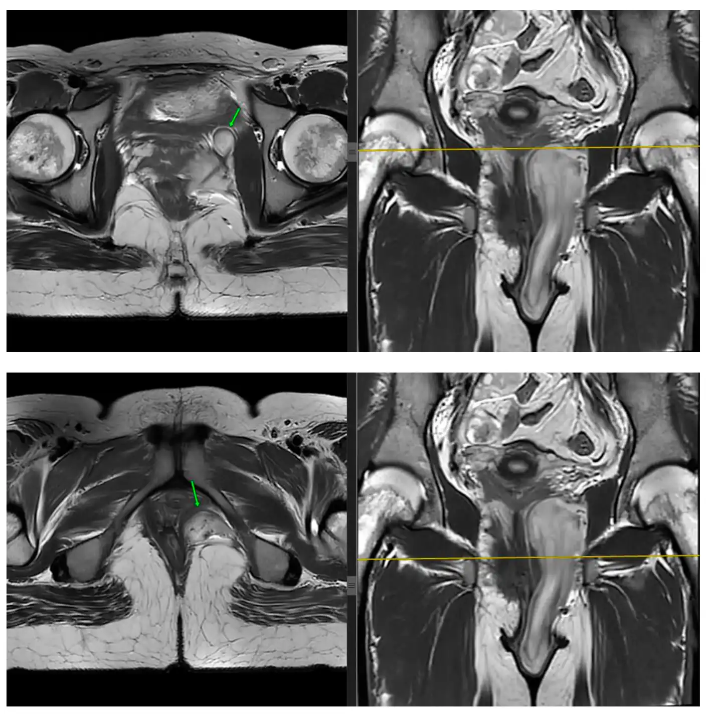
MRI reported a locally aggressive lesion involving the left labia majora, labia minora, supra-levator space, infra-levator space and left ischiorectal fossa. Based on imaging, the differential diagnosis included:
⦿ During surgery, a 10 × 5 × 4 cm pelvic mass was identified between the vagina, rectum and levator muscle. The tumour was adherent to the obturator internus muscle, while the rectum remained free, and the cervix and vagina appeared grossly normal.
⦿ Tumours arising in the perineum, ischiorectal fossa and pelvic floor are technically challenging because they lie close to important pelvic muscles, nerves and organs.
⦿ Successful surgery requires:
Pelvic soft tissue tumours should ideally be managed by surgeons experienced in complex pelvic surgery rather than treated as routine soft tissue swellings.
⦿ Although MRI strongly suggested Aggressive Angiomyxoma, the final diagnosis was established only after complete surgical excision.
⦿ Histopathology demonstrated an infiltrative tumour composed of loosely arranged spindle to stellate-shaped cells embedded in myxoid stroma with dilated blood vessels. Importantly, there was no nuclear atypia, no significant mitoses and no necrosis. The final diagnosis was Deep Aggressive Angiomyxoma.
⦿ This case highlights an important principle in pelvic oncology:
⦿ MRI suggests the diagnosis. Histopathology confirms it.
⦿ Many rare pelvic soft tissue tumours have overlapping imaging appearances. Definitive diagnosis should always be based on tissue examination after expert pathological assessment.
⦿ Histopathology showed a perineal mass measuring 11 × 6 × 4 cm, containing a 10.5 × 5 × 3 cm tumour with a shiny mucoid cut surface. Most importantly, the resection margin was free of tumour.
⦿ Deep Aggressive Angiomyxoma is a locally infiltrative tumour. Although it rarely spreads to distant organs, it has a well-recognised tendency for local recurrence if residual tumour is left behind.
Therefore, treatment should focus on:
Margin-negative surgery remains one of the most important predictors of durable local control.
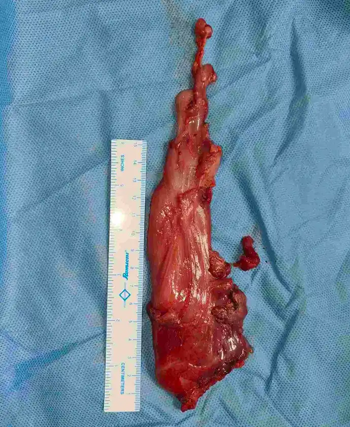
Following excision of this Deep Aggressive Angiomyxoma, the patient recovered well.
⦿ Hemodynamically stable
⦿ Ambulatory
⦿ Surgical wound clean
⦿ Pain well controlled
⦿ Drain placed safely in the pelvis
⦿ Discharged in stable condition with follow-up advice
Even after technically demanding pelvic surgery involving the levator muscles, vagina and perineal compartment, recovery can be smooth with protocol-based perioperative care, meticulous surgery, early mobilisation, good pain control and close postoperative monitoring.
Patients are often surprised that recovery after major pelvic tumour surgery is much better than expected when managed in experienced centres.
⦿ This patient’s journey demonstrates the complete management of a rare pelvic tumour.
⦿ MRI identified a lesion involving the left labia majora, labia minora, ischiorectal fossa, supra-levator and infra-levator space, suspicious for Aggressive Angiomyxoma. Surgery revealed a 10 × 5 × 4 cm pelvic mass between the vagina, rectum and levator muscle, which was removed intact. Histopathology confirmed Deep Aggressive Angiomyxoma, and importantly, all surgical margins were free of tumour.
⦿ Deep Aggressive Angiomyxoma is uncommon, but because it can extend through multiple pelvic compartments, treatment requires:
Patients searching for Deep Aggressive Angiomyxoma treatment, aggressive angiomyxoma surgery, perineal tumour surgery, pelvic soft tissue tumour specialist, labial tumour surgery, ischiorectal fossa tumour or pelvic mass excision should be evaluated by a surgeon experienced in complex pelvic and perineal tumour surgery.
⦿ Age alone does not determine whether adrenal tumour surgery is possible. The decision depends on the patient’s physiological reserve, comorbidities, tumour characteristics, functional status and expected benefit from surgery.
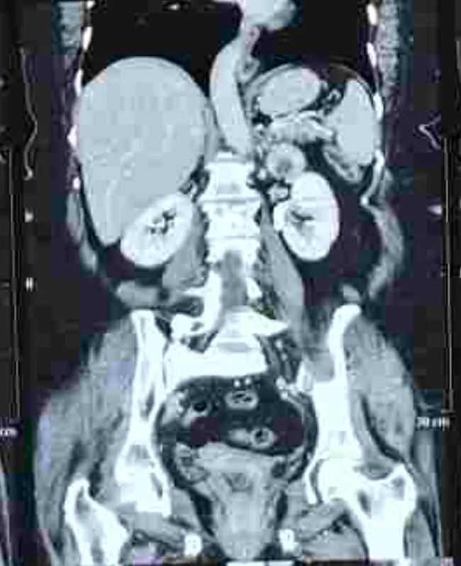
⦿ In this 71-year-old woman, imaging identified a 4 × 3.3 cm heterogeneous left suprarenal mass. The CT also documented significant associated findings, including focal fibrotic interstitial lung abnormality, emphysematous changes and mosaic attenuation, making comprehensive preoperative evaluation especially relevant.
⦿ Cardiopulmonary fitness
⦿ Anaesthetic risk
⦿ Functional status
⦿ Hormonal evaluation of an adrenal mass
⦿ Imaging characteristics and suspicion of malignancy
⦿ Expected oncological benefit
Despite her age and comorbidities, she underwent robotic adrenalectomy.
The key message is that elderly age or multiple comorbidities do not automatically rule out adrenal tumour surgery. With appropriate selection and perioperative planning, minimally invasive robotic surgery can be considered in suitable patients.
Not every adrenal mass is malignant. Imaging assessment is an important part of deciding whether observation, further evaluation or surgery is appropriate.
In this case, CT showed a well-defined, rounded, heterogeneous attenuation lesion measuring 4 × 3.3 cm in the left suprarenal fossa. The report recommended contrast-enhanced CT (CECT) for further evaluation.
When evaluating an adrenal mass, clinicians look at:
⦿ The patient’s clinical condition and endocrine evaluation are also important.
⦿ When imaging and clinical assessment raise sufficient concern for malignancy, adrenalectomy provides definitive pathological diagnosis as well as treatment.
⦿ In this patient, the adrenal lesion was ultimately managed surgically with robotic adrenalectomy.
⦿ The adrenal glands are located deep in the upper abdomen, close to major blood vessels, kidneys and other important structures. Precise dissection is therefore important, particularly when the diagnosis is uncertain or malignancy is being considered.
⦿ In this 71-year-old patient, imaging demonstrated a 4 × 3.3 cm heterogeneous left suprarenal mass. Because the patient also had multiple comorbidities, achieving the required oncological treatment while limiting surgical stress was an important consideration.
⦿ Magnified three-dimensional surgical vision
⦿ Articulating instruments for precise dissection
⦿ Improved access to a deep anatomical location
⦿ Minimally invasive access in appropriately selected patients
⦿ Potential for less postoperative morbidity compared with open surgery in suitable cases
The choice of approach must still depend on tumour size, imaging characteristics, suspicion of malignancy, local invasion and surgeon expertise. Open surgery may be more appropriate for large or invasive adrenal malignancies.
In this case, the patient underwent robotic adrenalectomy despite advanced age and multiple comorbidities.
⦿ Recently managed case by Dr Swati Shah – Robotic Uro-Oncology & Pelvic Oncosurgeon of a young patient with a history of obturator fossa synovial sarcoma, who developed recurrence at the previous surgical scar site.
⦿ The recurrence was identified as three nodules within the scar region over the lower back/flank. At surgery, a small approximately 3 × 2 cm mass with three nodules was found in the subcutaneous plane at the previous scar. There was no obvious disease elsewhere in the operated field.
⦿ The important point in recurrent soft-tissue sarcoma is that a new nodule or lump developing near a previous tumour or surgical site should not automatically be assumed to be scar tissue. Local recurrence of synovial sarcoma can present as a small mass or nodule, and timely assessment is important.
⦿ In this case, the recurrent mass was treated with wide local excision, with the mass removed intact along with an adequate margin.
⦿ Recently managed case by Dr Swati Shah – Robotic Uro-Oncology & Pelvic Oncosurgeon involving recurrent synovial sarcoma at a previous scar site.
⦿ The patient had a known history of obturator fossa synovial sarcoma. Clinical and pathological assessment showed recurrence at the lower-back/flank scar, described as three nodules.
⦿ During wide local excision, the operative finding was a small mass approximately 3 × 2 cm with three nodules within the previous scar, located in the subcutaneous plane. No other obvious disease was seen in the operative field.
⦿ This illustrates an important principle for patients who have previously undergone treatment for sarcoma: a new lump, nodule, swelling or change around the previous surgical scar deserves evaluation, even when the lesion appears small.
⦿ The lesion was excised intact with adequate margins and sent for histopathological examination.
⦿ Recently managed case by Dr Swati Shah – Robotic Uro-Oncology & Pelvic Oncosurgeon for recurrent synovial sarcoma at a previous scar site.
⦿ The operation performed was wide local excision of scar nodules with local flap reconstruction. Intraoperatively, the recurrent mass was dissected intact circumferentially, taking an adequate margin of the underlying muscle layer, rather than simply removing the visible nodules.
⦿ This approach is important in soft-tissue sarcoma because the objective is not merely to remove the palpable lump. The tumour needs to be removed with an appropriate oncological margin while preserving function and achieving safe wound closure.
⦿ The final histopathology in this case confirmed high-grade sarcoma consistent with synovial sarcoma, FNCLCC Grade III. The tumour measured 4 × 3 × 2 cm, and the closest surgical margin was 0.8 cm. Importantly, all examined surgical margins, including the base and skin margins, were free of tumour.
⦿ Thus, the operative plan and final pathology both demonstrate why wide local excision and adequate margins are central to surgery for recurrent soft-tissue sarcoma.
⦿ Recently managed case by Dr Swati Shah – Robotic Uro-Oncology & Pelvic Oncosurgeon of recurrent synovial sarcoma involving a previous lower-back/flank scar.
⦿ The excised specimen consisted of skin with underlying soft tissue measuring 6 × 4 × 3 cm. A circumscribed nodular tumour measuring 4 × 3 × 2 cm was identified. Histopathology showed:
⦿ The histopathology therefore provides more than just the diagnosis. Tumour grade, mitotic activity, necrosis and margin status are important components of the final oncological assessment.
⦿ In this patient, achieving tumour-free surgical margins after excision of the recurrent scar-site lesion was an important part of the surgical treatment.
⦿ Recently managed case by Dr Swati Shah – Robotic Uro-Oncology & Pelvic Oncosurgeon of recurrent synovial sarcoma at a previous lower-back/flank scar.
⦿ The final histopathology showed high-grade sarcoma, consistent with synovial sarcoma, FNCLCC Grade III. The report documented 18–19 mitoses per 10 high-power fields and necrosis of less than 5%.
⦿ FNCLCC grading is an important part of soft-tissue sarcoma pathology because it helps describe the biological aggressiveness of the tumour. The pathology report should therefore be reviewed along with the clinical history, imaging and surgical findings rather than considering the tumour size alone.
⦿ In this case, the excised tumour measured 4 × 3 × 2 cm, and importantly, all surgical margins, including the base and skin margins, were free of tumour.
⦿ Recently managed case by Dr Swati Shah – Robotic Uro-Oncology & Pelvic Oncosurgeon involving recurrence of obturator fossa synovial sarcoma at a previous scar site.
⦿ The recurrence appeared as three nodules within the previous lower-back/flank scar. At surgery, the lesion was found in the subcutaneous plane, with no obvious disease elsewhere in the operated field.
⦿ The recurrent tumour was treated by wide local excision, with the mass dissected intact circumferentially and an adequate margin of the underlying muscle layer taken with it.
⦿ The final pathology confirmed recurrent high-grade synovial sarcoma, FNCLCC Grade III, while demonstrating tumour-free surgical margins.
⦿ This case highlights why treatment of recurrent soft-tissue sarcoma requires careful assessment of the previous operative site and an oncological surgical plan rather than simple removal of a visible scar nodule.
⦿ Recently managed case by Dr Swati Shah – Robotic Uro-Oncology & Pelvic Oncosurgeon for recurrent synovial sarcoma involving a previous scar over the lower back/flank.
⦿ The operation performed was wide local excision of scar nodules with local flap reconstruction.
⦿ The recurrent mass was approximately 3 × 2 cm with three nodules in the subcutaneous plane. An elliptical incision was made including the overlying skin, and the tumour was dissected intact with an adequate margin of the muscle layer. After tumour removal, haemostasis was achieved, a drain was placed and the wound was closed in layers with local tissue management.
⦿ The final pathology showed a 4 × 3 × 2 cm synovial sarcoma, with the closest surgical margin measuring 0.8 cm and all examined margins free of tumour.
⦿ This demonstrates how surgery for recurrent soft-tissue sarcoma may require both oncological tumour removal and planned reconstruction to achieve safe closure of the surgical defect.
⦿ Recently managed case by Dr Swati Shah – Robotic Uro-Oncology & Pelvic Oncosurgeon involving a young patient with a history of obturator fossa synovial sarcoma who subsequently developed recurrence at the previous surgical scar.
⦿ The recurrence was clinically important despite being a relatively small lesion: the operative findings showed three nodules within the previous scar, with a mass approximately 3 × 2 cm in the subcutaneous plane.
⦿ The recurrent lesion was treated with wide local excision, and histopathology confirmed high-grade synovial sarcoma, FNCLCC Grade III, with 18–19 mitoses/10 HPF. All surgical margins were free of tumour.
⦿ A previous history of sarcoma therefore remains relevant even when a patient appears well or a new lesion near the scar is small. Long-term surveillance and prompt assessment of new nodules or swellings around a previous tumour site are important parts of sarcoma care.
Swasth Parivar: twice a month, practical advice on recovery, diet and warning signs. No advertising. Stop any time.
Your number is used for these updates and for the clinic to reach you. It is never sold or passed on. To stop, reply STOP on WhatsApp.