TURBT stands for Transurethral Resection of Bladder Tumour. It is a procedure used to examine, remove and obtain tissue from a suspicious growth inside the urinary bladder. The instrument is passed through the natural urinary passage (urethra), so there is no incision on the abdomen.
TURBT is often the first surgical procedure when bladder cancer is suspected. It helps establish the diagnosis, determine how deeply the tumour has grown and guide the next step in treatment.
TURBT has two important purposes: diagnosis and treatment. The removed tissue is examined by a pathologist to confirm the type of tumour, its grade and its stage.
Where technically safe and feasible, the surgeon aims to remove all visible tumour. The procedure also helps determine whether the cancer is non-muscle-invasive or has reached the bladder muscle. This distinction can substantially change the treatment plan.
TURBT is usually performed under general or spinal anaesthesia. A resectoscope is passed through the urethra into the bladder. The surgeon examines the bladder lining, identifies the tumour and removes it using a suitable instrument, such as an electrical loop or another resection technique.
Tissue may be collected from the tumour and its base. The samples are sent for histopathology. The operative record should document the tumour’s size, number and location, whether removal appears complete, and any relevant findings or complications.
A urinary catheter is commonly placed at the end of the procedure. In some patients, bladder irrigation is used temporarily to help prevent blood clots from blocking urine flow.
Your treating team will review your symptoms, urine tests, cystoscopy findings and relevant imaging. Tell the team about all medicines, especially blood thinners, allergies, previous anaesthesia problems and other medical conditions.
Follow the hospital’s instructions about fasting and whether medicines should be paused or continued. Do not stop prescribed blood-thinning medicines on your own. The plan depends on your health, medicines and the reason for treatment.
Some blood in the urine, urinary frequency, urgency or a burning sensation may occur for a short period. These symptoms should gradually improve. The catheter may be removed when the urine is sufficiently clear and the treating team considers it safe; the timing varies from patient to patient.
Recovery depends on the size and number of tumours, the depth and extent of resection, other medical conditions and whether further treatment is needed. Follow the discharge instructions about fluids, activity, medicines and follow-up. Avoid strenuous activity until your treating team advises that it is safe.
Contact your hospital urgently if you cannot pass urine, develop heavy or increasing bleeding or large clots, have fever or chills, experience worsening abdominal pain, or feel markedly unwell. Follow the emergency instructions provided at discharge.
The pathology report is essential for planning treatment. Important details include the tumour type, grade, stage, whether the tumour has invaded the lamina propria or bladder muscle, whether detrusor muscle is present in the specimen, and whether carcinoma in situ or other higher-risk features are identified.
Grade describes how abnormal the cancer cells look under the microscope. Stage describes how far the tumour has grown into the bladder wall or beyond it. Grade and stage are different, and both matter.
If the report does not include bladder muscle, this does not automatically mean the procedure was unsuccessful. However, depending on the tumour’s grade and stage, another resection may be recommended to improve staging and check for residual disease.
Yes. A repeat TURBT may be advised if the first resection was incomplete or its completeness is uncertain, if the tumour is T1, or in selected high-grade tumours when bladder muscle is absent from the specimen. When indicated, it is commonly planned within about two to six weeks, based on the clinical situation and recovery.
A second TURBT is not required for every patient. The decision depends on the initial operative findings, histopathology and individual risk.
For many patients with non-muscle-invasive bladder cancer, TURBT is the first and most important treatment step. Some patients may also need a medicine placed directly into the bladder, such as intravesical chemotherapy or BCG, depending on their risk group.
If the tumour has invaded the bladder muscle or has other high-risk features, TURBT alone may not be sufficient. Further staging and discussion of treatments such as systemic therapy, radical cystectomy or selected bladder-preserving approaches may be necessary.
Potential risks include bleeding, blood clots, urinary infection, difficulty passing urine after catheter removal, injury or perforation of the bladder, and the need for another procedure. The likelihood depends on the tumour and the patient’s health. Your surgeon and anaesthesia team can explain the risks relevant to your case.
Dr. Swati H. Shah is a uro- and gynaecologic oncosurgeon at Apollo Hospital, Ahmedabad, and Shah’s Cancer & Robotic Surgery Centre, Gota. Her bladder cancer practice includes TURBT, repeat TURBT when indicated, interpretation of pathology in the context of the operative findings, and planning further treatment for patients who require it.
The appropriate treatment depends on the individual diagnosis, tumour characteristics, general health and patient preferences. A consultation can help clarify the purpose of TURBT, the expected recovery and the next steps after the pathology report.
No. The instrument is passed through the urethra, so there is usually no abdominal incision.
A catheter is commonly used temporarily after TURBT. How long it stays depends on bleeding, the extent of resection and recovery.
The duration varies with the number, size and location of tumours and the complexity of the procedure.
The timing varies by laboratory and the tests required. Your treating team will explain the report and its implications.
Yes. Recurrence is possible, so follow-up cystoscopy and other treatment may be needed according to the tumour’s risk category.
No. It is recommended for specific findings, such as an incomplete resection, T1 disease or selected high-grade tumours without muscle in the sample.
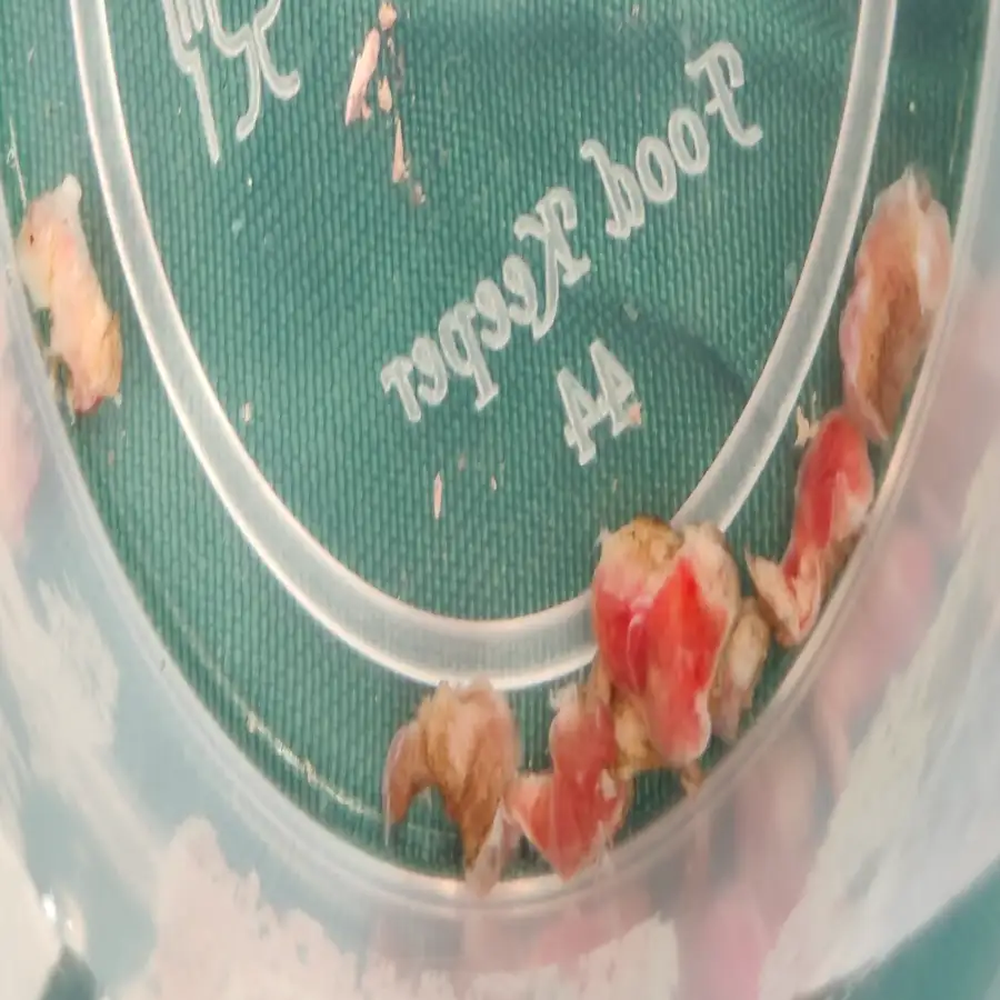
A 69-year-old male presented with intermittent, visible blood in his urine, a classic warning sign of underlying bladder pathology. Diagnostic imaging identified a 2 cm mass located on the right lateral wall, necessitating a precise intervention to both treat the lesion and obtain a definitive tissue diagnosis for staging.
⦿ Transurethral Resection of Bladder Tumor (TURBT)
• Procedure: Performed a complete endoscopic resection of the 2 cm right lateral bladder wall tumor.
• Staging Accuracy: Captured a targeted deep muscle biopsy to accurately determine the depth of tumor invasion.
• Safety / Blood Loss: Achieved successful intraoperative hemostasis, ensuring the patient remained hemodynamically stable throughout the procedure.
• Functional Preservation: Integrated a Double J stent placement to safeguard kidney function and maintain smooth urinary flow.
⦿ Rapid Recovery and Early Ambulation
The patient’s recovery was immediate and uneventful, with pain levels expertly managed through minimally invasive protocols. He was fully ambulatory and ready for discharge within just 24 hours of the procedure, returning home in stable condition. This proactive surgical intervention provided both a functional cure for the hematuria and a critical pathological roadmap for his future surveillance.
Visible blood in urine is a signal that demands immediate action. Through this minimally invasive endoscopic approach, we were able to completely remove the tumor and provide accurate staging, allowing this patient to return to his daily life almost immediately while securing a clear path for long-term monitoring.
This video features a patient testimonial and educational insight from Dr. Swati Shah, a renowned Robotic Uro-Oncologist and Gynecologic Cancer Surgeon. The primary focus of the video is on “Robotic Cystectomy with Neobladder,” a highly advanced surgical procedure used to treat urinary bladder cancer. When a patient’s urinary bladder is entirely removed due to cancer (a procedure called a cystectomy), a major concern is how they will pass urine afterward. The video addresses this critical question by explaining the concept of a “neobladder”—a new bladder surgically created from the patient’s own intestine, allowing them to urinate naturally without the need for an external stoma bag.
The video features a heartwarming and positive patient testimonial dedicated to Dr. Swati Shah, a highly regarded Robotic Uro-Oncologist and Gynecological Cancer Surgeon. It highlights the successful treatment of an elderly patient, who traveled from Kotvada village in the Bhavnagar district of Gujarat to seek specialized medical care. He recently underwent a complex Radical Cystectomy—a major surgical procedure to remove the urinary bladder—for the treatment of urinary bladder cancer under the expert supervision of Dr. Shah at Apollo Hospital. In the brief clip, the patient’s nephew shares their medical journey, explaining that they discovered Dr. Swati Shah’s profile online while searching for the best possible urological cancer treatment.
This video features a heartfelt patient testimonial detailing a successful bladder cancer surgery performed by Dr. Swati Shah, a highly skilled Robotic Uro-Oncologist. In the clip, a patient shares their personal recovery journey following a complex Robotic Radical Cystectomy, a major procedure where a new urinary bladder, known as a neobladder, was surgically created using advanced robotic technology. The patient explains that they underwent this operation two months prior. Although they initially felt anxious and fearful about the potential complications of such a significant medical procedure, the modern robotic approach and Dr. Shah’s expertise made the experience remarkably smooth.
This page provides general educational information and does not replace an individual medical consultation. The recommended procedure and follow-up depend on the patient’s clinical findings and histopathology.
Swasth Parivar: every week, practical advice on recovery, diet and warning signs. No advertising. Stop any time.
Your number is used for these updates and for the clinic to reach you. It is never sold or passed on. To stop, reply STOP on WhatsApp.